Copyright
©The Author(s) 2015.
World J Gastroenterol. Jul 7, 2015; 21(25): 7921-7928
Published online Jul 7, 2015. doi: 10.3748/wjg.v21.i25.7921
Published online Jul 7, 2015. doi: 10.3748/wjg.v21.i25.7921
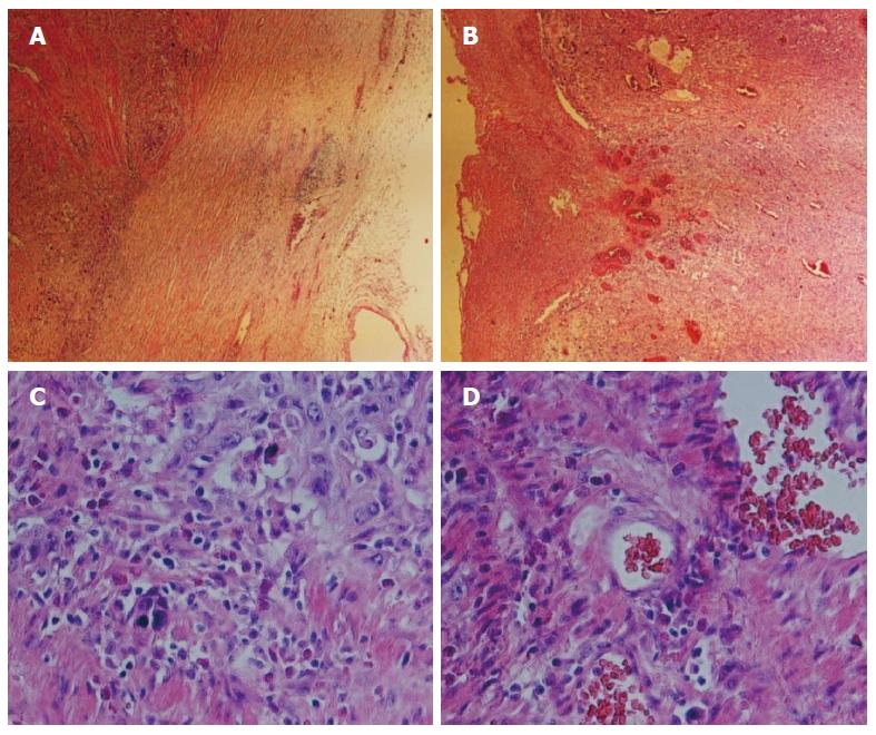
Figure 1 Histopathologic characterization of the esophageal squamous cell carcinoma.
Hematoxylin and eosin staining of the patient’s pathological tissue slide. A and B: Histologic sections revealed a papillary architecture (magnification × 40); C and D: Higher-magnification views of the slides. The tissue presents structural disorder involving abnormal organization, heterotypic cell number, deep nuclear staining, loss of normal epithelial polar structure, and increased mitotic activity. Obvious tumor nests are shown in (D) (magnification × 200).
- Citation: Qiao YY, Lin KX, Zhang Z, Zhang DJ, Shi CH, Xiong M, Qu XH, Zhao XH. Monitoring disease progression and treatment efficacy with circulating tumor cells in esophageal squamous cell carcinoma: A case report. World J Gastroenterol 2015; 21(25): 7921-7928
- URL: https://www.wjgnet.com/1007-9327/full/v21/i25/7921.htm
- DOI: https://dx.doi.org/10.3748/wjg.v21.i25.7921


