Copyright
©The Author(s) 2015.
World J Gastroenterol. May 7, 2015; 21(17): 5432-5441
Published online May 7, 2015. doi: 10.3748/wjg.v21.i17.5432
Published online May 7, 2015. doi: 10.3748/wjg.v21.i17.5432
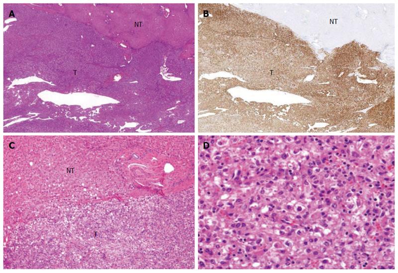
Figure 4 Microscopic features of the operative specimen.
A and C: Show low and high power views, respectively, of the borderline area of the tumor (T) and non-tumor (NT) areas (magnification × 20 and × 200, respectively, hematoxylin eosin staining); B: Shows immunostaining features using an anti-HMB45 antibody (magnification × 20); D: Shows the tumor including fat (magnification × 200; hematoxylin eosin staining).
- Citation: Maebayashi T, Abe K, Aizawa T, Sakaguchi M, Ishibashi N, Abe O, Takayama T, Nakayama H, Matsuoka S, Nirei K, Nakamura H, Ogawa M, Sugitani M. Improving recognition of hepatic perivascular epithelioid cell tumor: Case report and literature review. World J Gastroenterol 2015; 21(17): 5432-5441
- URL: https://www.wjgnet.com/1007-9327/full/v21/i17/5432.htm
- DOI: https://dx.doi.org/10.3748/wjg.v21.i17.5432


