Copyright
©The Author(s) 2015.
World J Gastroenterol. Mar 21, 2015; 21(11): 3394-3401
Published online Mar 21, 2015. doi: 10.3748/wjg.v21.i11.3394
Published online Mar 21, 2015. doi: 10.3748/wjg.v21.i11.3394
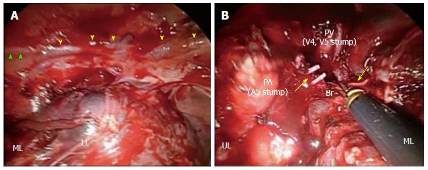
Figure 4 Intraoperative findings.
A: The presence of a collateral bronchial artery (BA) arising from the right inferior phrenic artery running along the phrenic nerve (green arrowheads) was confirmed; the tissue was black in color due to the first bronchial arterial embolization procedure (yellow arrowheads). B: Engorged right BAs streaming into the right middle lobe were dissected at the level of bifurcation of the middle lobe bronchus (arrows). UL: Upper lobe; ML: Middle lobe; LL: Lower lobe; Br: Middle lobe bronchus; PA: Pulmonary artery; PV: Pulmonary vein.
- Citation: Kitajima T, Momose K, Lee S, Haruta S, Ueno M, Shinohara H, Fujimori S, Fujii T, Takei R, Kohno T, Udagawa H. Bronchial bleeding caused by recurrent pneumonia after radical esophagectomy for esophageal cancer. World J Gastroenterol 2015; 21(11): 3394-3401
- URL: https://www.wjgnet.com/1007-9327/full/v21/i11/3394.htm
- DOI: https://dx.doi.org/10.3748/wjg.v21.i11.3394


