Copyright
©The Author(s) 2015.
World J Gastroenterol. Mar 21, 2015; 21(11): 3394-3401
Published online Mar 21, 2015. doi: 10.3748/wjg.v21.i11.3394
Published online Mar 21, 2015. doi: 10.3748/wjg.v21.i11.3394
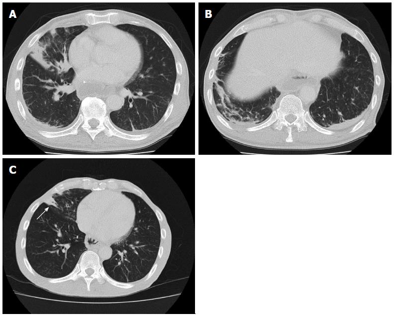
Figure 1 Unenhanced computed tomography after esophagectomy.
A: Computed tomography (CT) revealed consolidation in the right middle lobe; B: CT simultaneously demonstrated consolidation in the right lower lobe (A and B); C: Although there was no evidence of newly developed pneumonia, consolidation remained in the right middle lobe 14 mo after esophagectomy (arrow).
- Citation: Kitajima T, Momose K, Lee S, Haruta S, Ueno M, Shinohara H, Fujimori S, Fujii T, Takei R, Kohno T, Udagawa H. Bronchial bleeding caused by recurrent pneumonia after radical esophagectomy for esophageal cancer. World J Gastroenterol 2015; 21(11): 3394-3401
- URL: https://www.wjgnet.com/1007-9327/full/v21/i11/3394.htm
- DOI: https://dx.doi.org/10.3748/wjg.v21.i11.3394


