Copyright
©2014 Baishideng Publishing Group Inc.
World J Gastroenterol. Nov 14, 2014; 20(42): 15549-15563
Published online Nov 14, 2014. doi: 10.3748/wjg.v20.i42.15549
Published online Nov 14, 2014. doi: 10.3748/wjg.v20.i42.15549
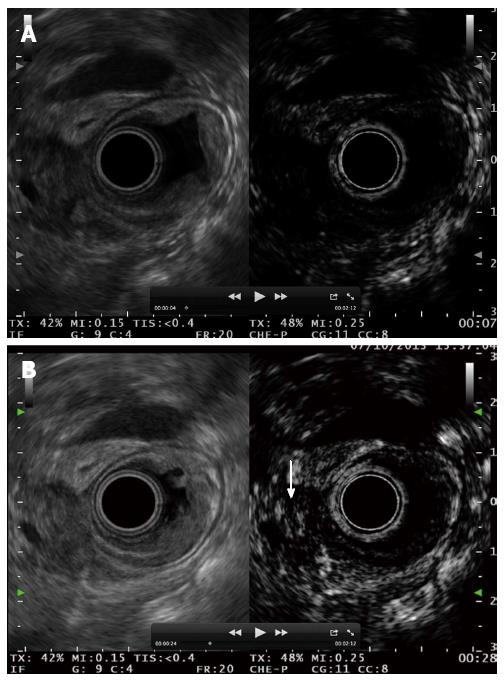
Figure 13 Ampullary tumour with echoic material inside the distal bile duct.
A: No enhancement is observed 7 s after contrast injection; B: Bile duct involvement is diagnosed after contrast enhancement 28 s after the injection.
- Citation: Alvarez-Sánchez MV, Napoléon B. Contrast-enhanced harmonic endoscopic ultrasound imaging: Basic principles, present situation and future perspectives. World J Gastroenterol 2014; 20(42): 15549-15563
- URL: https://www.wjgnet.com/1007-9327/full/v20/i42/15549.htm
- DOI: https://dx.doi.org/10.3748/wjg.v20.i42.15549


