Copyright
©2014 Baishideng Publishing Group Inc.
World J Gastroenterol. Sep 21, 2014; 20(35): 12687-12690
Published online Sep 21, 2014. doi: 10.3748/wjg.v20.i35.12687
Published online Sep 21, 2014. doi: 10.3748/wjg.v20.i35.12687
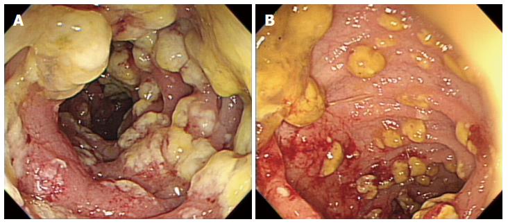
Figure 3 Follow-up sigmoidoscopy.
A: Sigmoidoscopy shows multiple yellowish plaques in the sigmoid colon. B: Sigmoidoscopy shows focal hemorrhagic changes in the sigmoid colon.
-
Citation: Kim JE, Gweon TG, Yeo CD, Cho YS, Kim GJ, Kim JY, Kim JW, Kim H, Lee HW, Lim T, Ham H, Oh HJ, Lee Y, Byeon J, Park SS. A case of
Clostridium difficile infection complicated by acute respiratory distress syndrome treated with fecal microbiota transplantation. World J Gastroenterol 2014; 20(35): 12687-12690 - URL: https://www.wjgnet.com/1007-9327/full/v20/i35/12687.htm
- DOI: https://dx.doi.org/10.3748/wjg.v20.i35.12687


