Copyright
©2013 Baishideng Publishing Group Co.
World J Gastroenterol. Dec 21, 2013; 19(47): 9133-9136
Published online Dec 21, 2013. doi: 10.3748/wjg.v19.i47.9133
Published online Dec 21, 2013. doi: 10.3748/wjg.v19.i47.9133
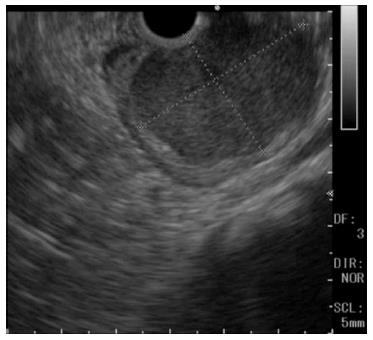
Figure 2 Findings on upper endoscopic ultrasonography.
A homogeneous, hypoechoic, well-demarcated mass, approximately 4 cm in diameter with a flat border, arising from the fourth layer of the gastric wall.
- Citation: Wada T, Tanabe S, Ishido K, Higuchi K, Sasaki T, Katada C, Azuma M, Naruke A, Kim M, Koizumi W, Mikami T. DOG1 is useful for diagnosis of KIT-negative gastrointestinal stromal tumor of stomach. World J Gastroenterol 2013; 19(47): 9133-9136
- URL: https://www.wjgnet.com/1007-9327/full/v19/i47/9133.htm
- DOI: https://dx.doi.org/10.3748/wjg.v19.i47.9133


