Copyright
©2013 Baishideng Publishing Group Co.
World J Gastroenterol. Dec 21, 2013; 19(47): 9003-9011
Published online Dec 21, 2013. doi: 10.3748/wjg.v19.i47.9003
Published online Dec 21, 2013. doi: 10.3748/wjg.v19.i47.9003
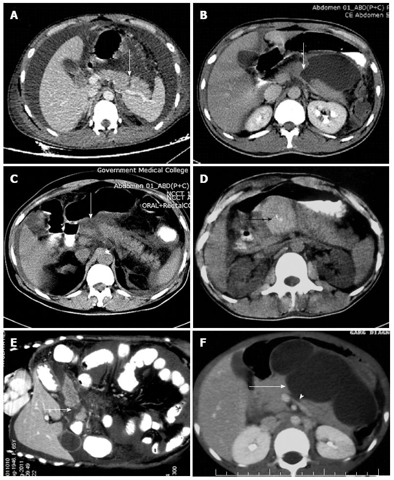
Figure 3 Computed tomography images.
A, B: Axial contrast-enhanced computed tomography shows a heterogeneous appearance of the body and tail of pancreas with a linear laceration (white arrow) across the distal body of the pancreas. There is also fluid in the lesser sac, perihepatic space, perisplenic space and hemoperitoneum. There is free air into chest wall muscles on right side in a case of blunt pancreatic trauma (A), and transection throughout extent of pancreatic parenchyma in proximal body region (suggestive of ductal injury) with a large fluid collection (white arrow) anterior to pancreas communication with the transection in another case of blunt injury to upper abdomen (B); C: Contrast-enhanced computed tomography demonstrating mild diffuse hypodensity of the body of pancreas. Contusions of the head and neck also demonstrated (white arrow) with secondary signs of traumatic pancreatitis, i.e., increased density of the peripancreatic fat, thickening of left anterior pararenal fascia, fluid in the lesser sac and hemoperitoneum; D: Plain axial computed tomography section at the level of pancreas shows a large hyperdense hematoma (black arrow) in proximal body of pancreas suggestive of pancreatic injury. E: Multiplanar reconstruction image of contrast-enhanced computed tomography demonstrating a pancreatic fracture (white arrow) in neck region with separation of pancreatic fragments; F: Contrast-enhanced axial computed tomography scan in a child with bicycle handlebar injury more than a month old shows a large lobulated pseudocyst anterior to pancreas communicating with pancreatic laceration in the neck of pancreas representing ductal injury. There is fluid between posterior pancreas and the splenic vein (arrow heads).
- Citation: Debi U, Kaur R, Prasad KK, Sinha SK, Sinha A, Singh K. Pancreatic trauma: A concise review. World J Gastroenterol 2013; 19(47): 9003-9011
- URL: https://www.wjgnet.com/1007-9327/full/v19/i47/9003.htm
- DOI: https://dx.doi.org/10.3748/wjg.v19.i47.9003


