Copyright
©2013 Baishideng Publishing Group Co.
World J Gastroenterol. Aug 14, 2013; 19(30): 4907-4916
Published online Aug 14, 2013. doi: 10.3748/wjg.v19.i30.4907
Published online Aug 14, 2013. doi: 10.3748/wjg.v19.i30.4907
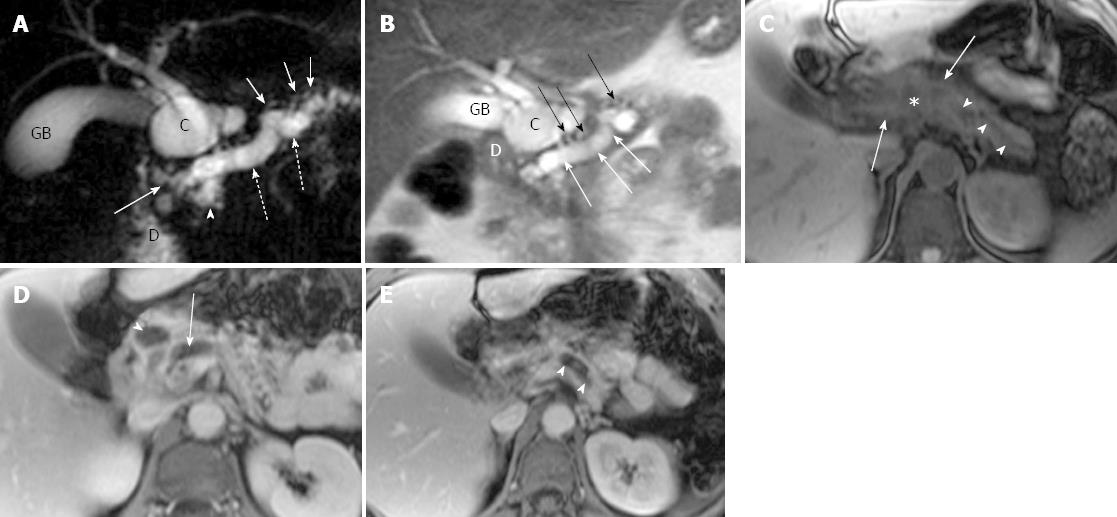
Figure 1 Magnetic resonance cholangiopancreatography and magnetic resonance imaging of recurrent acute pancreatitis involving the entire pancreas in a 44-year-old woman with several episodes of abdominal pain.
A: Coronal oblique thick-section rapid acquisition with relaxation enhancement magnetic resonance (RARE-MR) cholangiogram [infinite/1100 (effective), 40-mm section thickness] shows severe dilatation of pancreatic duct (dotted arrows) with stricture (solid arrow) just before entering the duodenum (D) and side branch ectasia (short arrows) as well. There is a pseudocyst (C) formation in pancreatic parenchyma. The gallbladder (GB) is distended and the common bile duct is dilated (arrowhead) as well; B: Thin-section half-Fourier RARE-MR cholangiogram [infinite/95 (effective), 3-mm section thickness] shows remarkable dilatation of dorsal pancreatic duct (white arrows) with severe side branch ectasia (black arrows). The pseudocyst (C) is formed in the pancreatic head region immediately adjacent to the duodenum (D) and GB; C: Axial precontrast T1WI SPGR shows the dilatation of pancreatic duct (arrowheads) and the pseudocyst (star) formation in the pancreatic neck were not easily appreciated. The signal intensity of the dorsal pancreas (arrows) is dramatically decreased; D: Axial postcontrast T1WI SPGR shows delayed enhancement of the dorsal pancreas and the wall of the pseudocysts (arrowhead). Dilatation of the pancreatic duct (arrow) was noted; E: Axial postcontrast T1WI SPGR shows delayed enhancement of the dorsal pancreas and the dilatation of the pancreatic duct (arrowheads).
- Citation: Wang DB, Yu J, Fulcher AS, Turner MA. Pancreatitis in patients with pancreas divisum: Imaging features at MRI and MRCP. World J Gastroenterol 2013; 19(30): 4907-4916
- URL: https://www.wjgnet.com/1007-9327/full/v19/i30/4907.htm
- DOI: https://dx.doi.org/10.3748/wjg.v19.i30.4907


