Copyright
©2012 Baishideng Publishing Group Co.
World J Gastroenterol. Aug 21, 2012; 18(31): 4102-4117
Published online Aug 21, 2012. doi: 10.3748/wjg.v18.i31.4102
Published online Aug 21, 2012. doi: 10.3748/wjg.v18.i31.4102
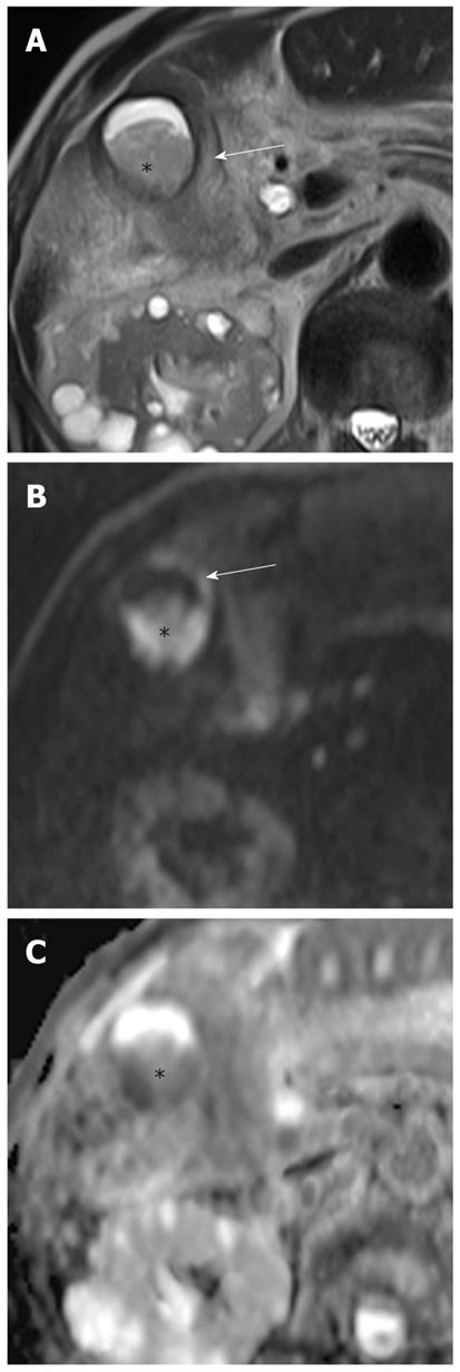
Figure 7 Gallbladder empyema in a 92-year-old man.
A: Axial T2-weighted rapid acquisition relaxation enhancement image demonstrates a fluid-fluid level with low signal intensity in the dependent portion (asterisk) of an inflamed gallbladder (arrow); B: DWI at b = 1000 s/mm2 shows purulent bile with high signal intensity (asterisk), and diffuse, symmetric high signal intensity in the wall of the gallbladder (arrow); C: On an ADC map, pus in the dependent portion of the gallbladder appears with low signal intensity (asterisk) due to restriction of diffusion. Empyema was confirmed by aspiration of pus during percutaneous cholecystostomy. DWI: Diffusion-weighted magnetic resonance imaging; ADC: Apparent diffusion coefficient.
- Citation: Lee NK, Kim S, Kim GH, Kim DU, Seo HI, Kim TU, Kang DH, Jang HJ. Diffusion-weighted imaging of biliopancreatic disorders: Correlation with conventional magnetic resonance imaging. World J Gastroenterol 2012; 18(31): 4102-4117
- URL: https://www.wjgnet.com/1007-9327/full/v18/i31/4102.htm
- DOI: https://dx.doi.org/10.3748/wjg.v18.i31.4102


