Case Report Open Access
Copyright ©2012 Baishideng Publishing Group Co., Limited. All rights reserved.
World J Gastroenterol. Jul 28, 2012; 18(28): 3765-3769
Published online Jul 28, 2012. doi: 10.3748/wjg.v18.i28.3765
Direct cholangioscopy combined with double-balloon enteroscope-assisted endoscopic retrograde cholangiopancreatography
Tatsuya Koshitani, Shogo Matsuda, Koji Takai, Takayuki Motoyoshi, Makoto Nishikata, Yasuhide Yamashita, Toshihiko Kirishima, Naomi Yoshinami, Hiroyuki Shintani, Department of Gastroenterology, Kyoto City Hospital, Kyoto 6048845, Japan
Toshikazu Yoshikawa, Department of Molecular Gastroenterology and Hepatology, Kyoto Prefectural University of Medicine Graduate School of Medical Science, Kyoto 6028566, Japan
Author contributions: Koshitani T and Matsuda S performed the research; Takai K, Motoyoshi T, Nishikata M, Yamashita Y, Kirishima T and Yoshinami N assisted with the research; Shintani H and Yoshikawa T supervised the research; Koshitani T and Matsuda S analyzed the data; Koshitani T wrote the paper.
Correspondence to: Tatsuya Koshitani, MD, Department of Gastroenterology, Kyoto City Hospital, 1-2 Mibu Higashitakada-cho, Nakagyo-ku, Kyoto 6048845, Japan. tkoshitani@aol.com
Telephone: +81-75-3115311 Fax: +81-75-3216025
Received: August 8, 2011
Revised: November 21, 2011
Accepted: April 22, 2012
Published online: July 28, 2012

Abstract

Double-balloon enteroscope (DBE)-assisted endoscopic retrograde cholangiopancreatography (ERCP) is an effective endoscopic approach for pancreatobiliary disorders in patients with altered gastrointestinal anatomy. Endoscopic interventions via DBE in these postoperative settings remain difficult because of the lack of an elevator and the use of extra-long ERCP accessories. Here, we report the usefulness of direct cholangioscopy with an ultra-slim gastroscope during DBE-assisted ERCP. Three patients with choledocholithiasis in postoperative settings (two patients after Billroth II gastrojejunostomy and one patient after Roux-en-Y gastrojejunostomy) were treated. DBE was used to gain access to the papilla under carbon dioxide insufflation, and endoscopic sphincterotomy was performed with a conventional sphincterotome. For direct cholangioscopy, the enteroscope was exchanged for an ultra-slim gastroscope through an incision in the overtube, which was inserted directly into the bile duct. Direct cholangioscopy was used to extract retained bile duct stones in two cases and to confirm the complete clearance of stones in one case. Bile duct stones were eliminated with a 5-Fr basket catheter under direct visual control. No adverse events were noted in any of the three cases. Direct cholangioscopy with an ultra-slim gastroscope facilitates subsequent treatment within the bile duct. This procedure represents another potential option during DBE-assisted ERCP.

Key Words: Direct cholangioscopy, Double-balloon enteroscope, Endoscopic retrograde cholangiopancreatography


INTRODUCTION

Endoscopic retrograde cholangiopancreatography (ERCP)-related maneuvers are difficult in patients with altered gastrointestinal (GI) anatomy due to previous abdominal surgery. ERCP success rates using conventional methods in patients after Billroth II gastrojejunostomy, Whipple pancreatoduodenectomy and Roux-en-Y gastrojejunostomy are reported to be as low as 52%[1,2], 51%[3], and 33%[2], respectively. If the papilla or biliary anastomosis in the afferent loop cannot be reached by an ordinary duodenoscope because of excessive intestinal length and/or sharp angulation of the anastomosis, it has recently become possible to use a double-balloon enteroscope (DBE) for ERCP. There are several hurdles for this technique, including reaching the papilla or biliary anastomosis, selective duct cannulation, and accomplishment of the treatment. The enteroscope is advanced through the overtube into the afferent jejunal loop using the push-and-pull double balloon technique. Identifying the opening of the afferent loop and coping with sharp angulation of the anastomosis are the keys for the successful endoscope insertion. Once the papilla or biliary anastomosis is reached, however, endoscopic interventions via DBE in these postoperative settings remain the most difficult ERCP manipulations because of the lack of an elevator and the use of extra-long ERCP accessories.

Here, we report three cases with choledocholithiasis treated with DBE-assisted ERCP and demonstrate the usefulness of direct cholangioscopy with an ultra-slim gastroscope for subsequent treatment.

CASE REPORT
Case 1

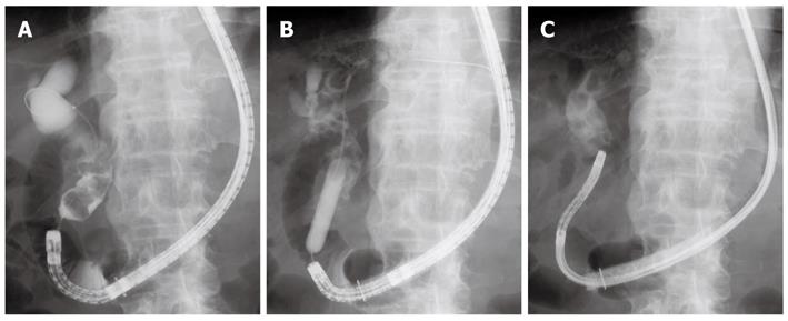
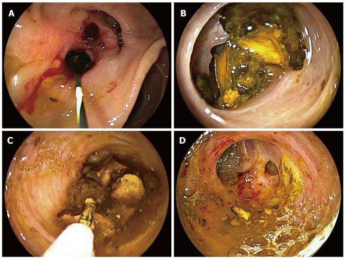
An 80-year-old man was referred for the treatment of choledocholithiasis. He had undergone partial gastrectomy with Roux-en-Y gastrojejunostomy for gastric carcinoma five years before. With the DBE (EC-450BI5; Fujinon, Osaka, Japan) with a 152 cm working length and a 2.8 mm instrumental channel, the papilla could be reached via the Roux-en-Y anastomosis under carbon dioxide insufflation. Cannulation of the papilla and cholangiography revealed a dilated bile duct and multiple stones (maximum size 20 mm) piled up at the lower to middle part of the bile duct (Figure 1A). Endoscopic sphincterotomy (EST) was performed with a conventional sphincterotome, placing the papilla of Vater at the 6 o’clock position. After mechanical lithotripsy with a 7-Fr crusher catheter (Xemex crusher catheter; Zeon Medical, Tokyo, Japan), stone extraction was attempted with a basket catheter but frequently failed because the catheter was inserted into the dilated cystic duct. Because the same attempt was performed unsuccessfully during another session, the enteroscope was exchanged for an ultra-slim gastroscope (EG-530NW; Fujinon, outer diameter 5.9 mm), leaving the overtube with its balloon inflated. The ultra-slim endoscope was introduced through an incision in the overtube, which was made at a point 100 cm from its tip. The ultra-slim endoscope was advanced through the overtube up to the papilla and was directly inserted into the bile duct after papillary dilation using a large balloon catheter (CRE balloon dilator; Boston Scientific Japan, Tokyo, Japan) (Figure 1B and C). Cholangioscopy showed multiple stones inside the dilated bile duct and the cystic duct. The stones were all removed with a 5-Fr basket catheter (Basket catheter, diamond type; MTW Endoskopie, Wesel, Germany) under direct visual control (Figure 2). The patient had a good clinical course after the procedure, and no adverse events occurred.

Figure 1
Figure 1 X-ray images. A: Cholangiography revealed a dilated bile duct and multiple stones piled up at the lower to middle part of the bile duct; B: Papillary dilation using a large balloon catheter; C: An ultra-slim gastroscope was introduced directly into the bile duct.
Figure 2
Figure 2 Endoscopic images. A: The papilla of Vater after balloon dilation; B: Cholangioscopy showed multiple stones inside the bile duct; C, D: The stones were all removed with a 5-Fr basket catheter under direct visual control.
Case 2

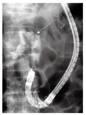
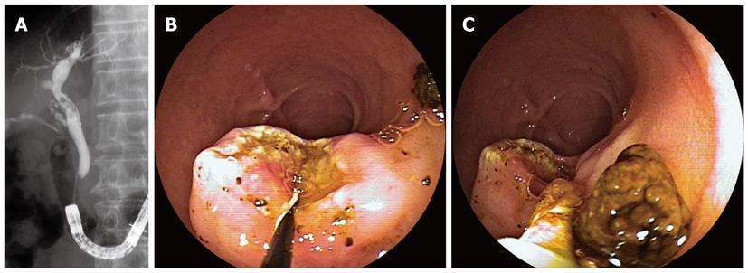
An 89-year-old man developed acute cholangitis with choledocholithiasis. He had undergone partial gastrectomy with Billroth II gastrojejunostomy for gastric carcinoma 21 years before. Because of the long afferent loop, the DBE (EC-450BI5; Fujinon, Osaka, Japan) was used to gain access to the papilla under carbon dioxide insufflation. Cannulation of the papilla and cholangiography showed two bile duct stones (maximum size 15 mm) at the middle part of the bile duct. After EST with a conventional sphincterotome, a smaller stone located distally was extracted with a basket catheter (Figure 3). Another stone could not be removed despite all possible efforts because of its impaction. The enteroscope was then exchanged for an ultra-slim gastroscope as described above. The endoscope was inserted directly into the bile duct, revealing an impacted stone inside the duct. The stone was removed successfully with a 5-Fr basket catheter under direct visual control, and the proximal bile duct was also investigated (Figure 4A-C). The patient had a good clinical course after the procedure, and no adverse events occurred.

Figure 3
Figure 3 Cholangiography. A smaller stone located distally was extracted with a basket catheter. Another stone was impacted inside the bile duct (arrow).
Figure 4
Figure 4 Direct cholangioscopy with an ultra-slim gastroscope. A: Cholangioscopy revealed an impacted stone inside the bile duct; B: The stone was removed with a 5-Fr basket catheter under direct visual control; C: The proximal bile duct was also investigated.
Case 3

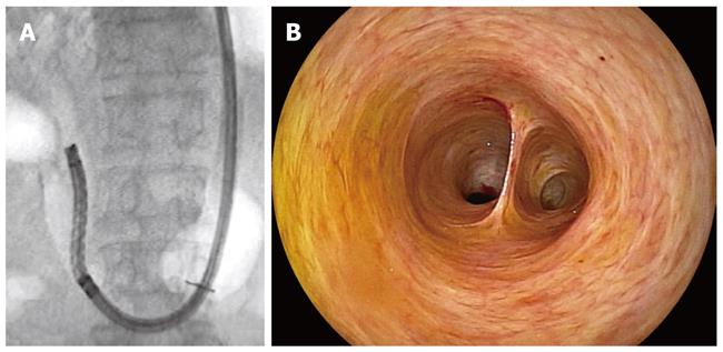
A 73-year-old man was referred because of cholestasis and suspected choledocholithiasis. He had undergone partial gastrectomy with Billroth II gastrojejunostomy for gastric ulcer 43 years before. The DBE (EC-450BI5; Fujinon, Osaka, Japan) was first inserted into the efferent loop and was then introduced up to the papilla via Braun’s anastomosis under carbon dioxide insufflation. Cannulation of the papilla and cholangiography revealed several small stones (5 mm in size) inside the bile duct. After EST with a conventional sphincterotome, the stones were extracted with a basket catheter and a retrieval balloon (Figure 5A-C). An ultra-slim gastroscope was then introduced into the bile duct as described above. Cholangioscopy confirmed complete clearance of the stones inside the bile duct (Figure 6A and B). The patient had a good clinical course after the procedure, and no adverse events occurred.

Figure 5
Figure 5 X-ray and endoscopic images. A: Cholangiography revealed several small stones inside the bile duct; B: The papilla of Vater after endoscopic sphincterotomy; C: The bile duct stones were extracted with a basket catheter and a retrieval balloon.
Figure 6
Figure 6 Direct cholangioscopy. A: An ultra-slim gastroscope was introduced directly into the bile duct after removal of bile duct stones; B: Cholangioscopy confirmed complete clearance of the duct.
DISCUSSION

Since the first report[4] of a successful DBE-assisted cholangiography in a pediatric case with late biliary stricture of choledochojejunostomy after living donor liver transplantation, many case series[5-17] have reported the effectiveness of DBE-assisted ERCP for pancreatobiliary disorders in various postoperative settings. In most previous reports[6-8,10-12,15,16], the therapeutic enteroscope, which has a 200-cm working length and a 2.8-mm instrumental channel, has been used for DBE-assisted ERCP. Recently, the single-balloon enteroscope (SBE) system (Olympus Medical Systems, Tokyo, Japan), which consists of an enteroscope without the balloon at the tip of the endoscope and an overtube equipped with a balloon, has also been used for ERCP in patients with altered postsurgical anatomy with similar technical success rates[18-20].

The maneuverability of these enteroscopes is rather poor and only limited ERCP accessories are available for these endoscopes because of the long working length. To overcome these drawbacks, Fähndrich et al[9] reported a facilitated method for endoscopic interventions at the bile duct during DBE-assisted ERCP that involved exchanging the enteroscope with an ordinary gastroscope after incision of the overtube once the papilla or the biliary anastomosis was reached. This technique is favorably performed in our country when the SBE or the “long” DBE is used for ERCP because long-length ERCP accessories are not commercially available in Japan. Alternatively, the DBE, which has a 152 cm working length and a 2.8 mm instrumental channel, can be used. This instrument is the “short” DBE for which conventional ERCP accessories are available. With this enteroscope, Shimatani et al[13] performed DBE-assisted ERCP and related therapeutic maneuvers in 68 patients with altered GI anatomies with an extremely high success rate.

Another disadvantage of DBE-assisted ERCP is that the enteroscope is forward-viewing. The lack of an elevator and the absence of the side-viewing perspective reduce the success rate of cannulation or sphincterotomy. Moreover, additional treatment becomes difficult in cases such as the retained bile duct stones described here, even if access to the biliary duct is obtained. Direct visualization of the bile duct with an ultra-slim gastroscope expands the options for therapy inside the duct because the endoscope has a 2 mm instrumental channel for which 5-Fr accessories (including the biopsy forceps, the basket catheter, the laser lithotripsy probe, and the argon plasma coagulation probe) are available. The ultra-slim endoscope was introduced through incision of the overtube. The overtube was incised for three-quarters of its circumference on the side opposite of the pressure line, as described by Fähndrich et al[9] Care was taken not to damage the pressure line of the overtube to maintain the inflation of the balloon. Although this technique is only possible when the intact papilla or biliary anastomosis is reached with (at most) 100 cm of the enteroscope introduced, it is easy to advance the ultra-slim endoscope into the bile duct through the papilla treated with EST or balloon dilation because the approach is from the distal side of the afferent loop.

We did not encounter any adverse events in this case series. However, care must be taken because DBE-assisted ERCP can potentially cause serious complications, such as perforation, bleeding, or pancreatitis, during or after the scope insertion and ERCP procedures. Although no serious complications of direct cholangioscopy with an ultra-slim endoscope have been reported[21,22], there is still a risk of air embolism; pneumobilia is often recognized after EST. Carbon dioxide insufflation during the DBE procedure may be effective in preventing this complication.

In conclusion, DBE-assisted ERCP appears to be a promising method; however, this technique still has several disadvantages. Direct cholangioscopy with an ultra-slim gastroscope compensates for drawbacks of the enteroscope and facilitates subsequent treatment within the bile duct. This procedure may represent another option during DBE-assisted ERCP.

Footnotes

Peer reviewer: Ibrahim A Al Mofleh, Professor, Department of Medicine, College of Medicine, King Saud University, PO Box 2925, Riyadh 11461, Saudi Arabia

S- Editor Cheng JX L- Editor A E- Editor Zheng XM

References
1.  Lin LF, Siauw CP, Ho KS, Tung JC. ERCP in post-Billroth II gastrectomy patients: emphasis on technique. Am J Gastroenterol. 1999;94:144-148.  [PubMed]  [DOI]  [Cited in This Article: ]  [Cited by in Crossref: 68]  [Cited by in F6Publishing: 74]  [Article Influence: 3.0]  [Reference Citation Analysis (0)]
2.  Hintze RE, Adler A, Veltzke W, Abou-Rebyeh H. Endoscopic access to the papilla of Vater for endoscopic retrograde cholangiopancreatography in patients with billroth II or Roux-en-Y gastrojejunostomy. Endoscopy. 1997;29:69-73.  [PubMed]  [DOI]  [Cited in This Article: ]  [Cited by in Crossref: 145]  [Cited by in F6Publishing: 158]  [Article Influence: 5.9]  [Reference Citation Analysis (0)]
3.  Chahal P, Baron TH, Topazian MD, Petersen BT, Levy MJ, Gostout CJ. Endoscopic retrograde cholangiopancreatography in post-Whipple patients. Endoscopy. 2006;38:1241-1245.  [PubMed]  [DOI]  [Cited in This Article: ]  [Cited by in Crossref: 102]  [Cited by in F6Publishing: 113]  [Article Influence: 6.3]  [Reference Citation Analysis (0)]
4.  Haruta H, Yamamoto H, Mizuta K, Kita Y, Uno T, Egami S, Hishikawa S, Sugano K, Kawarasaki H. A case of successful enteroscopic balloon dilation for late anastomotic stricture of choledochojejunostomy after living donor liver transplantation. Liver Transpl. 2005;11:1608-1610.  [PubMed]  [DOI]  [Cited in This Article: ]  [Cited by in Crossref: 95]  [Cited by in F6Publishing: 96]  [Article Influence: 5.1]  [Reference Citation Analysis (0)]
5.  Emmett DS, Mallat DB. Double-balloon ERCP in patients who have undergone Roux-en-Y surgery: a case series. Gastrointest Endosc. 2007;66:1038-1041.  [PubMed]  [DOI]  [Cited in This Article: ]  [Cited by in Crossref: 150]  [Cited by in F6Publishing: 160]  [Article Influence: 9.4]  [Reference Citation Analysis (0)]
6.  Aabakken L, Bretthauer M, Line PD. Double-balloon enteroscopy for endoscopic retrograde cholangiography in patients with a Roux-en-Y anastomosis. Endoscopy. 2007;39:1068-1071.  [PubMed]  [DOI]  [Cited in This Article: ]  [Cited by in Crossref: 146]  [Cited by in F6Publishing: 145]  [Article Influence: 8.5]  [Reference Citation Analysis (0)]
7.  Maaser C, Lenze F, Bokemeyer M, Ullerich H, Domagk D, Bruewer M, Luegering A, Domschke W, Kucharzik T. Double balloon enteroscopy: a useful tool for diagnostic and therapeutic procedures in the pancreaticobiliary system. Am J Gastroenterol. 2008;103:894-900.  [PubMed]  [DOI]  [Cited in This Article: ]  [Cited by in Crossref: 70]  [Cited by in F6Publishing: 74]  [Article Influence: 4.6]  [Reference Citation Analysis (0)]
8.  Chu YC, Yang CC, Yeh YH, Chen CH, Yueh SK. Double-balloon enteroscopy application in biliary tract disease-its therapeutic and diagnostic functions. Gastrointest Endosc. 2008;68:585-591.  [PubMed]  [DOI]  [Cited in This Article: ]  [Cited by in Crossref: 59]  [Cited by in F6Publishing: 68]  [Article Influence: 4.3]  [Reference Citation Analysis (0)]
9.  Fähndrich M, Sandmann M, Heike M. A facilitated method for endoscopic interventions at the bile duct after Roux-en-Y reconstruction using double balloon enteroscopy. Z Gastroenterol. 2008;46:335-338.  [PubMed]  [DOI]  [Cited in This Article: ]  [Cited by in Crossref: 11]  [Cited by in F6Publishing: 12]  [Article Influence: 0.8]  [Reference Citation Analysis (0)]
10.  Koornstra JJ, Fry L, Mönkemüller K. ERCP with the balloon-assisted enteroscopy technique: a systematic review. Dig Dis. 2008;26:324-329.  [PubMed]  [DOI]  [Cited in This Article: ]  [Cited by in Crossref: 39]  [Cited by in F6Publishing: 47]  [Article Influence: 3.1]  [Reference Citation Analysis (0)]
11.  Kuga R, Furuya CK, Hondo FY, Ide E, Ishioka S, Sakai P. ERCP using double-balloon enteroscopy in patients with Roux-en-Y anatomy. Dig Dis. 2008;26:330-335.  [PubMed]  [DOI]  [Cited in This Article: ]  [Cited by in Crossref: 22]  [Cited by in F6Publishing: 27]  [Article Influence: 1.8]  [Reference Citation Analysis (0)]
12.  Mönkemüller K, Fry LC, Bellutti M, Neumann H, Malfertheiner P. ERCP with the double balloon enteroscope in patients with Roux-en-Y anastomosis. Surg Endosc. 2009;23:1961-1967.  [PubMed]  [DOI]  [Cited in This Article: ]  [Cited by in Crossref: 49]  [Cited by in F6Publishing: 65]  [Article Influence: 4.1]  [Reference Citation Analysis (0)]
13.  Shimatani M, Matsushita M, Takaoka M, Koyabu M, Ikeura T, Kato K, Fukui T, Uchida K, Okazaki K. Effective "short" double-balloon enteroscope for diagnostic and therapeutic ERCP in patients with altered gastrointestinal anatomy: a large case series. Endoscopy. 2009;41:849-854.  [PubMed]  [DOI]  [Cited in This Article: ]  [Cited by in Crossref: 158]  [Cited by in F6Publishing: 170]  [Article Influence: 11.3]  [Reference Citation Analysis (0)]
14.  Ryozawa S, Iwamoto S, Iwano H, Ishigaki N, Taba K, Sakaida I. ERCP using double-balloon endoscopes in patients with Roux-en-Y anastomosis. J Hepatobiliary Pancreat Surg. 2009;16:613-617.  [PubMed]  [DOI]  [Cited in This Article: ]  [Cited by in Crossref: 16]  [Cited by in F6Publishing: 18]  [Article Influence: 1.2]  [Reference Citation Analysis (0)]
15.  Parlak E, Ciçek B, Dişibeyaz S, Cengiz C, Yurdakul M, Akdoğan M, Kiliç MZ, Saşmaz N, Cumhur T, Sahin B. Endoscopic retrograde cholangiography by double balloon enteroscopy in patients with Roux-en-Y hepaticojejunostomy. Surg Endosc. 2010;24:466-470.  [PubMed]  [DOI]  [Cited in This Article: ]  [Cited by in Crossref: 56]  [Cited by in F6Publishing: 53]  [Article Influence: 3.5]  [Reference Citation Analysis (0)]
16.  Lin CH, Tang JH, Cheng CL, Tsou YK, Cheng HT, Lee MH, Sung KF, Lee CS, Liu NJ. Double balloon endoscopy increases the ERCP success rate in patients with a history of Billroth II gastrectomy. World J Gastroenterol. 2010;16:4594-4598.  [PubMed]  [DOI]  [Cited in This Article: ]  [Cited by in CrossRef: 14]  [Cited by in F6Publishing: 16]  [Article Influence: 1.1]  [Reference Citation Analysis (0)]
17.  Itoi T, Ishii K, Sofuni A, Itokawa F, Tsuchiya T, Kurihara T, Tsuji S, Ikeuchi N, Fukuzawa K, Moriyasu F. Long- and short-type double-balloon enteroscopy-assisted therapeutic ERCP for intact papilla in patients with a Roux-en-Y anastomosis. Surg Endosc. 2011;25:713-721.  [PubMed]  [DOI]  [Cited in This Article: ]  [Cited by in Crossref: 71]  [Cited by in F6Publishing: 66]  [Article Influence: 4.7]  [Reference Citation Analysis (0)]
18.  Dellon ES, Kohn GP, Morgan DR, Grimm IS. Endoscopic retrograde cholangiopancreatography with single-balloon enteroscopy is feasible in patients with a prior Roux-en-Y anastomosis. Dig Dis Sci. 2009;54:1798-1803.  [PubMed]  [DOI]  [Cited in This Article: ]  [Cited by in Crossref: 59]  [Cited by in F6Publishing: 51]  [Article Influence: 3.4]  [Reference Citation Analysis (0)]
19.  Neumann H, Fry LC, Meyer F, Malfertheiner P, Monkemuller K. Endoscopic retrograde cholangiopancreatography using the single balloon enteroscope technique in patients with Roux-en-Y anastomosis. Digestion. 2009;80:52-57.  [PubMed]  [DOI]  [Cited in This Article: ]  [Cited by in Crossref: 57]  [Cited by in F6Publishing: 68]  [Article Influence: 4.5]  [Reference Citation Analysis (0)]
20.  Itoi T, Ishii K, Sofuni A, Itokawa F, Tsuchiya T, Kurihara T, Tsuji S, Ikeuchi N, Umeda J, Moriyasu F. Single-balloon enteroscopy-assisted ERCP in patients with Billroth II gastrectomy or Roux-en-Y anastomosis (with video). Am J Gastroenterol. 2010;105:93-99.  [PubMed]  [DOI]  [Cited in This Article: ]  [Cited by in Crossref: 129]  [Cited by in F6Publishing: 123]  [Article Influence: 8.8]  [Reference Citation Analysis (0)]
21.  Moon JH, Ko BM, Choi HJ, Hong SJ, Cheon YK, Cho YD, Lee JS, Lee MS, Shim CS. Intraductal balloon-guided direct peroral cholangioscopy with an ultraslim upper endoscope (with videos). Gastrointest Endosc. 2009;70:297-302.  [PubMed]  [DOI]  [Cited in This Article: ]  [Cited by in Crossref: 111]  [Cited by in F6Publishing: 122]  [Article Influence: 8.1]  [Reference Citation Analysis (0)]
22.  Tsou YK, Lin CH, Tang JH, Liu NJ, Cheng CL. Direct peroral cholangioscopy using an ultraslim endoscope and overtube balloon-assisted technique: a case series. Endoscopy. 2010;42:681-684.  [PubMed]  [DOI]  [Cited in This Article: ]  [Cited by in Crossref: 21]  [Cited by in F6Publishing: 21]  [Article Influence: 1.5]  [Reference Citation Analysis (0)]