Copyright
©2012 Baishideng Publishing Group Co.
World J Gastroenterol. Jun 28, 2012; 18(24): 3177-3180
Published online Jun 28, 2012. doi: 10.3748/wjg.v18.i24.3177
Published online Jun 28, 2012. doi: 10.3748/wjg.v18.i24.3177
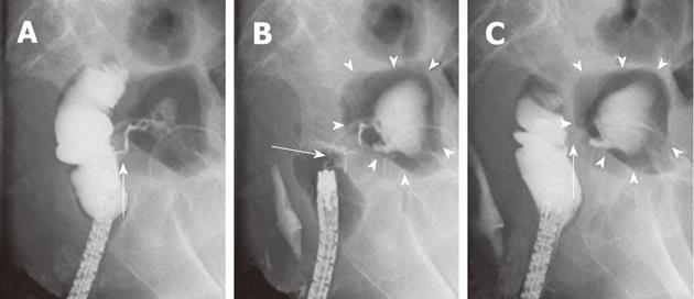
Figure 4 The contrast radiography of case 2.
A: The lateral view of the contrast radiography and the colonoscopy revealed a fistula communicating with the urinary bladder and the inflowing and pooling of the radiocontrast agent in the urinary bladder (arrow); B: The fistula was slightly larger than the single over-the-scope-clip (OTSC). Single OTSC closure (arrow) resulted in 1/3 of the fistula remaining. Again, the contrast radiography revealed a residual fistula leading to the urinary bladder (arrowheads); C: The remaining fistula was completely closed with double OTSCs. The contrast radiography revealed no fistula leading to the urinary bladder (arrowheads). Tests revealed the successful closure of the fistula without any inflowing of the radiocontrast agent (arrow).
- Citation: Mori H, Kobara H, Fujihara S, Nishiyama N, Kobayashi M, Masaki T, Izuishi K, Suzuki Y. Rectal perforations and fistulae secondary to a glycerin enema: Closure by over-the-scope-clip. World J Gastroenterol 2012; 18(24): 3177-3180
- URL: https://www.wjgnet.com/1007-9327/full/v18/i24/3177.htm
- DOI: https://dx.doi.org/10.3748/wjg.v18.i24.3177


