Copyright
©2012 Baishideng Publishing Group Co.
World J Gastroenterol. Mar 21, 2012; 18(11): 1273-1278
Published online Mar 21, 2012. doi: 10.3748/wjg.v18.i11.1273
Published online Mar 21, 2012. doi: 10.3748/wjg.v18.i11.1273
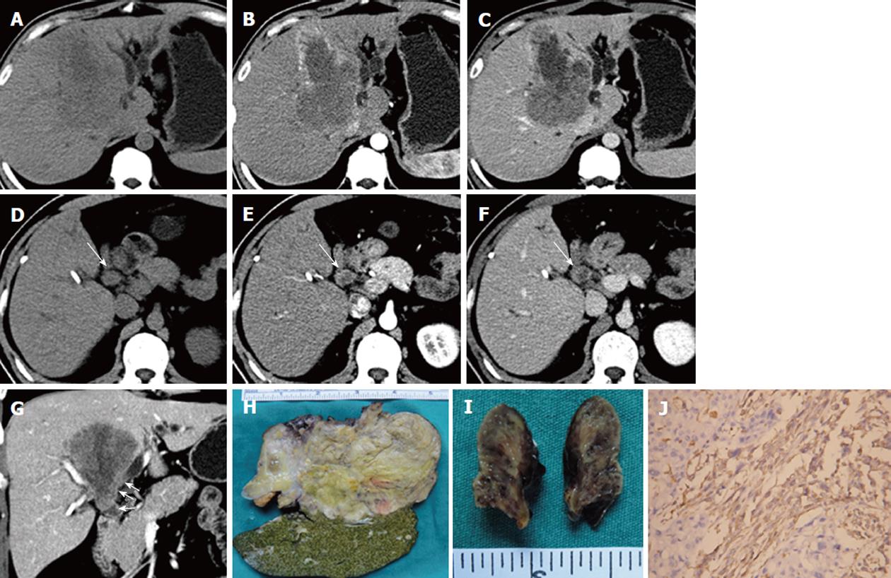
Figure 1 A 41-year-old man with primary hepatic carcinosarcoma.
A large hypodense tumor in the hepatic middle lobe appears on precontrast computed tomography (CT) scan (A), which shows early rim enhancement in the hepatic artery phase (B) and a low density in the portal phase with large areas of necrosis (C). The bile duct tumor thrombus (BDTT) (arrow) shows similar enhancement patterns with the intrahepatic tumor on pre-contrast CT scan (D), hepatic artery phase (E) and portal phase scan (F). Coronal reconstruction image in the portal phase shows that BDTT (arrows) is contiguous with the intrahepatic tumor (G). The resected hepatic tumor (H) and BDTT (I) specimens. The sarcomatous component of the primary hepatic carcinosarcoma is vimentin positive on the immunohistochemical staining (J), × 200.
- Citation: Liu QY, Lin XF, Li HG, Gao M, Zhang WD. Tumors with macroscopic bile duct thrombi in non-HCC patients: Dynamic multi-phase MSCT findings. World J Gastroenterol 2012; 18(11): 1273-1278
- URL: https://www.wjgnet.com/1007-9327/full/v18/i11/1273.htm
- DOI: https://dx.doi.org/10.3748/wjg.v18.i11.1273


