Copyright
©2011 Baishideng Publishing Group Co.
World J Gastroenterol. Sep 28, 2011; 17(36): 4090-4098
Published online Sep 28, 2011. doi: 10.3748/wjg.v17.i36.4090
Published online Sep 28, 2011. doi: 10.3748/wjg.v17.i36.4090
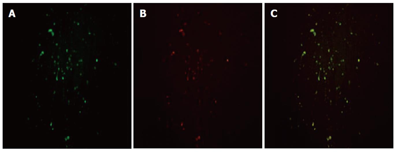
Figure 2 Double immunostaining was used to analyze the cellular localization of high-mobility group box 1 protein and α-smooth muscle actin in hepatic fibrosis tissue (original magnification, × 200).
A: α-smooth muscle actin (α-SMA) was stained with polyclonal α-SMA antibody and secondarily by rhodamine -conjugated anti-rabbit antibody (green); B: High-mobility group box 1 (HMGB1) was stained with monoclonal anti-HMGB1 antibody and secondarily by fluoresceinisothiocyanate-conjugated anti-rabbit antibody (red); C: The yellow areas on the merged image show co-localization of α-SMA and HMGB1.
- Citation: Ge WS, Wu JX, Fan JG, Wang YJ, Chen YW. Inhibition of high-mobility group box 1 expression by siRNA in rat hepatic stellate cells. World J Gastroenterol 2011; 17(36): 4090-4098
- URL: https://www.wjgnet.com/1007-9327/full/v17/i36/4090.htm
- DOI: https://dx.doi.org/10.3748/wjg.v17.i36.4090


