Copyright
©2011 Baishideng Publishing Group Co.
World J Gastroenterol. Apr 28, 2011; 17(16): 2086-2095
Published online Apr 28, 2011. doi: 10.3748/wjg.v17.i16.2086
Published online Apr 28, 2011. doi: 10.3748/wjg.v17.i16.2086
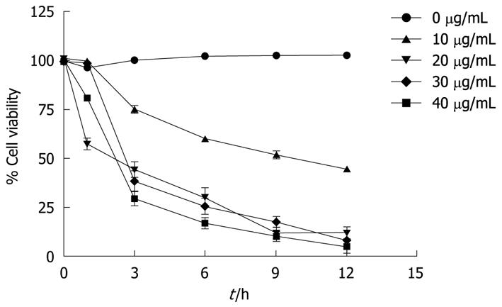
Figure 1 Effect of α-mangostin on the viability of COLO 205 cells.
Different concentrations of α-mangostin (0-40 μg/mL) at different incubation times were studied. Cell viability is expressed as a percentage of the number of viable cells to that of the control, to which no α-mangostin was applied. Each data point shown is the mean ± SD from three independent experiments.
- Citation: Watanapokasin R, Jarinthanan F, Nakamura Y, Sawasjirakij N, Jaratrungtawee A, Suksamrarn S. Effects of α-mangostin on apoptosis induction of human colon cancer. World J Gastroenterol 2011; 17(16): 2086-2095
- URL: https://www.wjgnet.com/1007-9327/full/v17/i16/2086.htm
- DOI: https://dx.doi.org/10.3748/wjg.v17.i16.2086


