Copyright
©2009 The WJG Press and Baishideng.
World J Gastroenterol. Dec 28, 2009; 15(48): 6126-6128
Published online Dec 28, 2009. doi: 10.3748/wjg.15.6126
Published online Dec 28, 2009. doi: 10.3748/wjg.15.6126
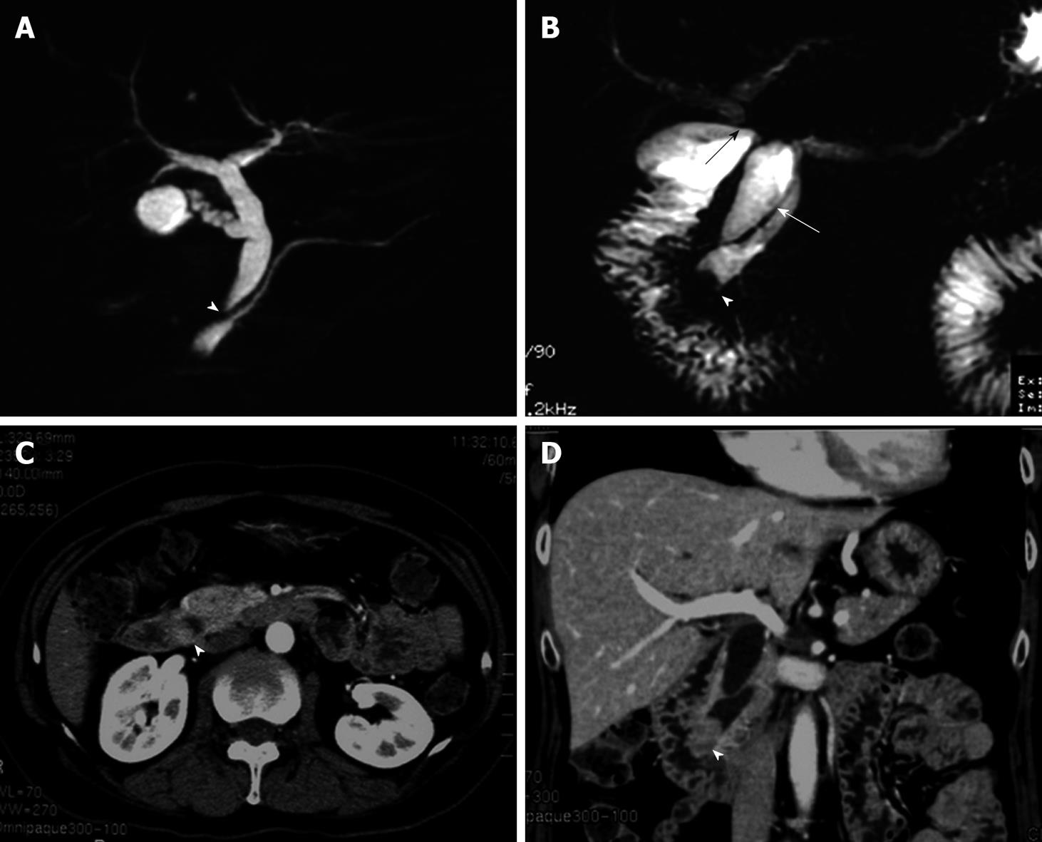
Figure 1 Imaging modalities for pancreaticobiliary maljunction and carcinoma of the papilla of Vater.
A: Magnetic resonance choledochopancreatography before first operation shows both maljunction of the main pancreatic duct and the choledochus (arrowhead) and the choledochal dilation, however there is no finding of neoplasm at the papilla of Vater; B: Magnetic resonance choledochopancreatography at follow-up after first operation shows the tumor in the main pancreatic duct (arrowhead). In addition, the choledochoduodenostomy (black arrow) and the residual choledochus (white arrow) can be seen; C, D: Computed tomography obtained 2 years after choledochoduodenostomy for pancreaticobiliary maljunction shows the mass in the distal end of the common bile duct, which had not been detected before first operation (arrowheads).
- Citation: Watanabe M, Midorikawa Y, Yamano T, Mushiake H, Fukuda N, Kirita T, Mizuguchi K, Sugiyama Y. Carcinoma of the papilla of Vater following treatment of pancreaticobiliary maljunction. World J Gastroenterol 2009; 15(48): 6126-6128
- URL: https://www.wjgnet.com/1007-9327/full/v15/i48/6126.htm
- DOI: https://dx.doi.org/10.3748/wjg.15.6126


