Copyright
©2009 The WJG Press and Baishideng.
World J Gastroenterol. Apr 14, 2009; 15(14): 1774-1778
Published online Apr 14, 2009. doi: 10.3748/wjg.15.1774
Published online Apr 14, 2009. doi: 10.3748/wjg.15.1774
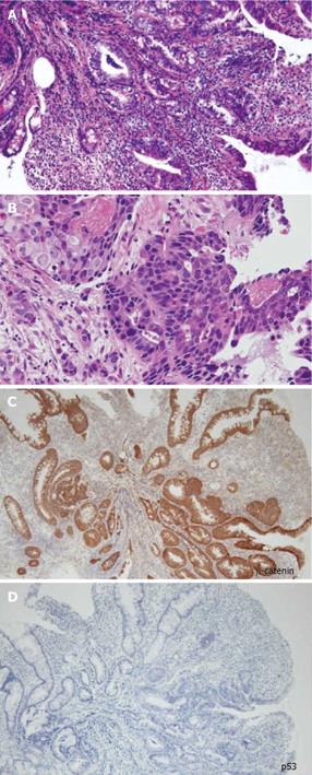
Figure 4 Histopathological findings of the biopsy specimen showed poorly differentiated adenocarcinoma.
A: HE (× 100), B: HE (× 400), C: Immunohistochemical staining using β-catenin antibody (× 100), D: Immunohistochemical staining using p53 antibody (× 100).
- Citation: Kodaira C, Osawa S, Mochizuki C, Sato Y, Nishino M, Yamada T, Takayanagi Y, Takagaki K, Sugimoto K, Kanaoka S, Furuta T, Ikuma M. A case of small bowel adenocarcinoma in a patient with Crohn’s disease detected by PET/CT and double-balloon enteroscopy. World J Gastroenterol 2009; 15(14): 1774-1778
- URL: https://www.wjgnet.com/1007-9327/full/v15/i14/1774.htm
- DOI: https://dx.doi.org/10.3748/wjg.15.1774


