Published online Oct 7, 2008. doi: 10.3748/wjg.14.5707
Revised: August 11, 2008
Accepted: August 18, 2008
Published online: October 7, 2008
AIM: To study the esophageal transit time (ETT) and compare its mean value among three anatomical inclinations of the body; and to analyze the correlation of ETT to body mass index (BMI).
METHODS: A biomagnetic technique was implemented to perform this study: (1) The transit time of a magnetic marker (MM) through the esophagus was measured using two fluxgate sensors placed over the chest of 14 healthy subjects; (2) the ETT was assessed in three anatomical positions (at upright, fowler, and supine positions; 90º, 45º and 0º, respectively).
RESULTS: ANOVA and Tuckey post-hoc tests demonstrated significant differences between ETT mean of the different positions. The ETT means were 5.2 ± 1.1 s, 6.1 ± 1.5 s, and 23.6 ± 9.2 s for 90º, 45º and 0º, respectively. Pearson correlation results were r = -0.716 and P < 0.001 by subjects’ anatomical position, and r = -0.024 and P > 0.05 according the subject’s BMI.
CONCLUSION: We demonstrated that using this biomagnetic technique, it is possible to measure the ETT and the effects of the anatomical position on the ETT.
- Citation: Cordova-Fraga T, Sosa M, Wiechers C, Roca-Chiapas JMDL, Moreles AM, Bernal-Alvarado J, Huerta-Franco R. Effects of anatomical position on esophageal transit time: A biomagnetic diagnostic technique. World J Gastroenterol 2008; 14(37): 5707-5711
- URL: https://www.wjgnet.com/1007-9327/full/v14/i37/5707.htm
- DOI: https://dx.doi.org/10.3748/wjg.14.5707
The esophageal phase is the last phase in the swallow process; it includes the propulsion of the meal through the esophagus toward the stomach. The esophageal transit time (ETT) reported for solid and semisolid meals is between 4 and 8 s, whereas liquid ETT lasts approximately 1 to 2 s in healthy people[1]. A diagnosis of gastroesophageal reflux disease should include the presence of a pathological reflux in patients lacking another motility disorder or damage in the esophagus[2,3]. If this condition can not be met, then the evaluation should include the assessment of disintegration time of oral tablets before they enter the stomach[4]. Currently, diagnosis of gastroesophageal reflux diseases is made with endoscopy[5,6], manometry[3], imaging methods[7], impedance[8], scintigraphy[9] and other techniques[2,10]. These assessments are useful to quantify the liquid and solid volumes retained in the esophagus. Currently, the scintigraphic technique is the gold standard test accepted to assess ETT and it is indicated in cases where the manometric and barometric studies do not give a differential diagnosis[11].
The ETT assessment is used to complete the diagnosis of diseases, such as gastroesophageal reflux[12-14], dysphagia[15,16], esophagitis[17,18], and achalasia[6,19,20]. The latter studies are commonly performed with scintigraphic and manometric techniques[21], in healthy[22], geriatric[23], and pediatric patients[24], despite the use of ionizing radiation and catheters in each test, respectively. Recently, several assessments were performed using the biomagnetic technique, including gastric emptying time[25] and colon transit time[26]. These studies have the advantage of being non-invasive, comfortable for the patient, and do not use ionizing radiation. Daghastanli et al[27] in 1998 reported an ETT study carried out with a biosusceptometer and a magnetic tracer, where they used 5 g of manganese (Mn) and ferrite power. In their study, they also measured the ETT using the scintigraphy technique and found that the ETT was 4.6 ± 0.9 s when biomagnetic technique was used, in comparison to a time of 3.8 ± 0.8 s as measured by the scintigraphy technique. The results of these studies demonstrated the efficacy of the magnetic techniques to carry out the ETT.
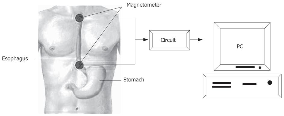
In this study, we implemented a novel modality of the biomagnetic technique using modern instruments, which included the monitoring of a magnetic marker (MM) traveling though the length of the esophagus. This was done using a pair of fluxgate magnetometers. We hypothesized that the esophagus motility and the mean ETT are significantly different when they are tested as a function of different anatomical positions of a subject (supine = 0°; fowler = 45°, and upright = 90°, respectively) (Figure 1).
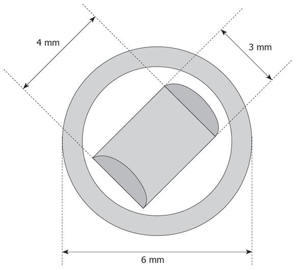
In Figure 1, we present a schematic set up of the biomagnetic probe used, which consisted of two digital tri-axis fluxgate magnetometers which were placed in an electronic device. They were separated 19 cm and were positioned in a line along the esophagus, just above the subjects’ thorax (Figure 2). A 3-mm long and 4-mm high magnet was used as the biomagnetic source or MM. This magnet was enclosed in a polycarbonate sphere 6 mm in diameter (Figure 3) in order to avoid chemical reactions with the gastric acids.
Fourteen normal and healthy adult subjects (10 men and 4 women) participated in the study; they did not have clinical antecedents of esophageal or gastrointestinal disease. The subject’s mean age and body mass index (BMI) were 21.8 ± 1.5 years and 23.9 ± 2.7 kg/m2, respectively. All volunteers received instructions before starting the experiment and signed an informed consent approved by the Institutional Review Board of our institution. The experiment was carried out according to the Declaration of Helsinki.
Subjects were studied after fasting for 12 h. All of them were assessed in three anatomical positions: (1) upright position, 90°; (2) fowler position, this is a semi-laying position bent at 45°; and (3) supine position, this is a laying position with a bend of 0° (Figure 1). The MM or magnetic particle was introduced inside the mouth of the subjects and swallowed with 20 mL of yogurt (50 kcal), this substance was used as MM vehicle.
The magnetic signal was registered for 1 min, with a sampling rate of 30 samples/s. Data acquisition was carried out using a routine informatic implemented with software of LabVIEW 7 platform. Then, collected data were exported and graphically analyzed in Matlab 6.5 in order to measure the ETT.
We calculated the mean age and BMI of the subjects using descriptive statistics. Using one way ANOVA and Tuckey post-hoc test, we compared the differences among the mean ETT obtained when subjects adopted each of the three anatomical positions. A Pearson correlation was used to determine the correlation coefficient between the ETT and the subject’s age, BMI and the angle of inclination of the anatomical position. P < 0.05 was considered statistically significant.
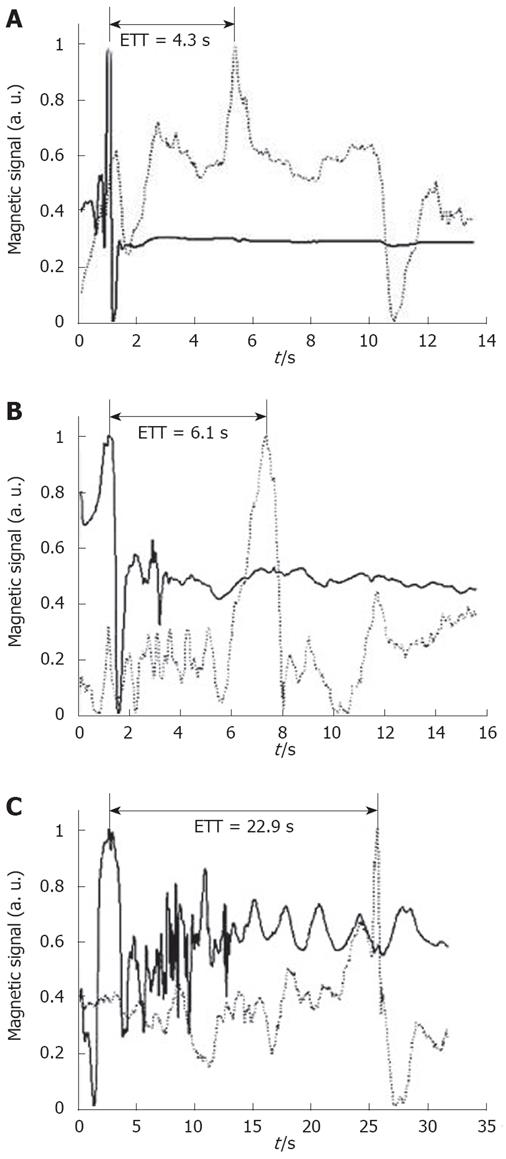
Figure 4 shows the raw signals recorded from one subject in three anatomical positions. The time signal shown as a continuous line is the recording with the fluxgate magnetometer added in the upper part of the esophagus, while the dashed line corresponds to the fluxgate magnetometer in the bottom part of the esophagus. The different time between the dominant peaks of each raw signal gives the ETT in each case. In Figure 4, it shows the raw recordings carried out in each anatomical position of one subject. We estimate that the differences in the time seen here was typical of all subjects.
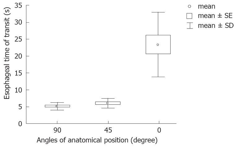
Figure 5 demonstrates the mean and standard deviation values of the ETTs, which were significantly longer at 0° (23.6 ± 9.2 s) than at 45° (6.1 ± 1.5 s), and 90° (5.18 ± 1.8 s). The results of the ANOVA and Tuckey post-hoc test demonstrated the significant differences between the groups (Figure 5). Pearson correlation coefficient test demonstrated an indirect relationship between anatomical position and ETT. This means that when subjects adopt a greater angle of inclination, they will have shorter ETT values. This relationship had a coefficient of r = -0.716, P < 0.001. However, we did not find any statistically significant difference between the EET and weight, age or BMI.
The esophageal phase is the last phase in the swallow process; it includes the propulsion of the meal through the esophagus toward the stomach. The ETT reported for solid and semisolid meals is between 4 and 8 s, whereas liquid ETT lasts approximately 1 to 2 s in healthy people[1]. Using the biomagnetic technique, we demonstrated that ETT is affected by anatomical position, with a significantly larger transit time when subjects adopted an upright position. These results concur with previous reports[1], including studies in which a biosusceptometer magnetometer was used[27].
Previously, researchers reported that ETT in healthy individuals was approximately 4-8 s for solid and semisolid meals, and 1-2 s for liquid meals[11,28,29]. In agreement with the aforementioned findings, our study demonstrated that the mean ETT was 6.1 ± 1.5 s in the upright position and 5.18 ± 1.8 s in the fowler position. However, the only value which was inconsistent with previous reports was that of the supine position, a transit time of 23.6 ± 9.2 s. This can be explained by the effects of gravity on the test meal and magnets.
Our study demonstrated that ETT is affected by gravity, and therefore, the subjects’ anatomical position changes ETT. This phenomenon is explained by the physiology of the esophagus, which combines resistance and contraction to cause movement of the bolus or liquid. When gravity also contributes to the propulsion of the bolus or liquid through the esophagus, transit time is decreased and esophageal transit rates are increased.
Using this biomagnetic modality, we demonstrated that ETT varies depending on the subjects’ anatomical position. In this study, we found no relationship between ETT and the subjects’ age, which can be explained by the fact that we generally evaluated only young and healthy subjects (mean age: 21.8 ± 1.5 years). However, more studies assessing the ETT in older subjects and patients with different pathologies, such as gastroesophageal reflux[12-14], dysphagia[15,16], esophagitis[17,18], and achalasia[6,19,20], are necessary to determine the differences in ETT of healthy subjects versus patients. It is likely that the ETT will differ in patients with a clinical diagnosis of esophageal disease.
In this study, we found no significant correlation between ETT and the subject’s BMI, which also may be explained by the samples from a largely homogeneous group of healthy and non-obese subjects (BMI: 23.9 ± 2.7 kg/m2). Therefore, in order to demonstrate the relationship of BMI and ETT, additional studies are needed using males and females with BMI within normal and obese ranges. Other clinical applications of biomagnetic technique exist in gastro-pharmacology studies, where researchers are testing the efficacy of a drug to increase or decrease ETT.
An advantage of this new application of the biomagnetic technique, implemented for the measurement of ETT, is that it demands little space and hardware, since all that is needed is a low-cost magnetometer. Therefore, this technique could be implemented for clinical assessment of esophageal disorders in general practice medicine, for gastroenterologists studying drug transit time[4,11] and in other specialties. Additionally, because of its low cost and non-invasiveness, this technique could be implemented in small clinical areas and hospitals. Although this technique has already been validated[27], further studies are needed to compare biomagnetism with the most innovative and sophisticated techniques commonly used for esophageal evaluation in order to identify its sensitivity and reproducibility.
Esophageal transit time (ETT) assessment is used to diagnose of gastroesophageal disease, such as esophagitis and achalasia. These studies are carried out using either scintigraphic or manometric techniques, but each has its own disadvantage: scintigraphy uses ionizing radiation, while manometry uses an invasive probe. Recently, several assessments of other body systems were performed using the biomagnetic technique (BMT), including ETT, gastric emptying time, and colon transit time. These studies have the advantage of being non-invasive, comfortable for the patient, and are conducted without ionizing radiation. Recently, researchers have tested the validity of BMT by comparing it with the scintigraphic technique (the gold standard test accepted to assess ETT) in the evaluation of ETT.
In this study, authors implemented a novel use of the BMT using modern instruments, which included the monitoring of a magnetic marker (MM) traveling though the length of the esophagus. Using the BMT technique, they demonstrated that ETT varies depending on the subjects’ anatomical position.
The Fluxgate magnetometer is a small device which functions at room temperature without a magnetic un-shielding room. Its range to precisely identify locations of a small MM is approximately a distance of less than 50 cm. It is possible to record magnetic signals through solid objects including the human body, and therefore, the technique is still valid if the MM is held in one's hand or is passing through the esophagus. Therefore, this is a portable evaluation system that can be implemented for the clinical study and diagnosis of esophegeal disorders in hospitals around the world.
A major application of this technique is the simultaneous collection of peristaltic activity data from different points along the esophagus. By a subject ingesting an MM similar in size and shape to a pill and following it as it passes through the esophageous by monitoring its progress at several sites along the esophagus, researchers can deduce the peristaltic behavior of the esophagus. Accordingly, ETT and propulsion velocity can accuratly be estimated using this technique.
Body anatomical inclinations is an angle of the human body, such as standing upright (90º), fowler (45º) and supine positions (0º). Biomagnetic technique is a technique which uses either external or internal magnetic fields applied to biological materials. Biomagnetism is the phenomenon that involves magnetic fields produced by the human body and other living entities. It is to be distinguished from magnetic fields applied to the body, properly called magnetobiology. Biosusceptometer magnetometer is a probe instrument using room-temperature sensor(s) that can measure variations in magnetic susceptibilities. MM means external magnetic substance or object used in this case as a vehicle to monitor the passage of an orally taken substance (food, drug, tablet, capsule, etc.) through the intestinal tract. Fluxgate sensor is a scientific instrument used to measure the strength and/or direction of the magnetic field in the vicinity of the instrument. Magnetic susceptibility means the magnetization of a material per unit applied field. It describes the magnetic response of a substance to an applied magnetic field.
This is an interesting study. Authors used a BMT to monitor ETT to test the hypothesis that esophageal motility and the mean ETT are significantly different when subjects adopt different anatomical inclinations. With this technique, they demonstrated that the mean values of ETT vary depending on the subjects’ anatomical inclination.
Peer reviewer: Michael F Vaezi, Department of Gastroentero-logy and Hepatology, Vanderbilt University Medical Center, 1501 TVC, Nashville, TN 37232-5280, United States
S- Editor Li DL L- Editor Kumar M E- Editor Lin YP
| 1. | Ojeaburu J, Benjamin SB. Normal Esophageal Fisiology. Gastrointestinal Disease an Endoscopic Approach. New Jersey: Slack Incorporated 2002; 173-190. [Cited in This Article: ] |
| 2. | Mariani G, Boni G, Barreca M, Bellini M, Fattori B, AlSharif A, Grosso M, Stasi C, Costa F, Anselmino M. Radionuclide gastroesophageal motor studies. J Nucl Med. 2004;45:1004-1028. [Cited in This Article: ] |
| 3. | Can MF, Yagci G, Cetiner S, Gulsen M, Yigit T, Ozturk E, Gorgulu S, Tufan T. Accurate positioning of the 24-hour pH monitoring catheter: agreement between manometry and pH step-up method in two patient positions. World J Gastroenterol. 2007;13:6197-6202. [Cited in This Article: ] |
| 4. | Perkins AC, Blackshaw PE, Hay PD, Lawes SC, Atherton CT, Dansereau RJ, Wagner LK, Schnell DJ, Spiller RC. Esophageal transit and in vivo disintegration of branded risedronate sodium tablets and two generic formulations of alendronic acid tablets: a single-center, single-blind, six-period crossover study in healthy female subjects. Clin Ther. 2008;30:834-844. [Cited in This Article: ] |
| 5. | Ahmadi A, Draganov P. Endoscopic mucosal resection in the upper gastrointestinal tract. World J Gastroenterol. 2008;14:1984-1989. [Cited in This Article: ] |
| 6. | Chung JJ, Park HJ, Yu JS, Hong YJ, Kim JH, Kim MJ, Lee SI. A comparison of esophagography and esophageal transit scintigraphy in the evaluation of usefulness of endoscopic pneumatic dilatation in achalasia. Acta Radiol. 2008;49:498-505. [Cited in This Article: ] |
| 7. | Gilja OH, Hatlebakk JG, Odegaard S, Berstad A, Viola I, Giertsen C, Hausken T, Gregersen H. Advanced imaging and visualization in gastrointestinal disorders. World J Gastroenterol. 2007;13:1408-1421. [Cited in This Article: ] |
| 8. | Blonski W, Hila A, Jain V, Freeman J, Vela M, Castell DO. Impedance manometry with viscous test solution increases detection of esophageal function defects compared to liquid swallows. Scand J Gastroenterol. 2007;42:917-922. [Cited in This Article: ] |
| 9. | Hike K, Urita Y, Watanabe T, Sugimoto M, Miki K. Saliva transit from the oral cavity to the esophagus in GERD. Hepatogastroenterology. 2008;55:4-7. [Cited in This Article: ] |
| 10. | Vergara MT. Labotarorio en los trastornos motores del esófago. Gastr Latinoam. 2005;16:109-114. [Cited in This Article: ] |
| 11. | Osmanoglou E, Van Der Voort IR, Fach K, Kosch O, Bach D, Hartmann V, Strenzke A, Weitschies W, Wiedenmann B, Trahms L. Oesophageal transport of solid dosage forms depends on body position, swallowing volume and pharyngeal propulsion velocity. Neurogastroenterol Motil. 2004;16:547-556. [Cited in This Article: ] |
| 12. | Sidhu SS, Bal C, Karak P, Garg PK, Bhargava DK. Effect of endoscopic variceal sclerotherapy on esophageal motor functions and gastroesophageal reflux. J Nucl Med. 1995;36:1363-1367. [Cited in This Article: ] |
| 13. | Tolin RD, Malmud LS, Reilley J, Fisher RS. Esophageal scintigraphy to quantitate esophageal transit (quantitation of esophageal transit). Gastroenterology. 1979;76:1402-1408. [Cited in This Article: ] |
| 14. | Stanghellini V, Tosetti C, Corinaldesi R. Standards for non-invasive methods for gastrointestinal motility: scintigraphy. A position statement from the Gruppo Italiano di Studio Motilita Apparato Digerente (GISMAD). Dig Liver Dis. 2000;32:447-452. [Cited in This Article: ] |
| 15. | Tatum RP, Shi G, Manka MA, Brasseur JG, Joehl RJ, Kahrilas PJ. Bolus transit assessed by an esophageal stress test in postfundoplication dysphagia. J Surg Res. 2000;91:56-60. [Cited in This Article: ] |
| 16. | Zhu ZJ, Chen LQ, Duranceau A. Long-term result of total versus partial fundoplication after esophagomyotomy for primary esophageal motor disorders. World J Surg. 2008;32:401-407. [Cited in This Article: ] |
| 17. | Dantas RO, Oliveira RB, Aprile LR, Hara SH, Sifrim DA. Saliva transport to the distal esophagus. Scand J Gastroenterol. 2005;40:1010-1016. [Cited in This Article: ] |
| 18. | De Jonge PJ, Van Eijck BC, Geldof H, Bekkering FC, Essink-Bot ML, Polinder S, Kuipers EJ, Siersema PD. Capsule endoscopy for the detection of oesophageal mucosal disorders: a comparison of two different ingestion protocols. Scand J Gastroenterol. 2008;43:870-877. [Cited in This Article: ] |
| 19. | Bonavina L, Nosadini A, Bardini R, Baessato M, Peracchia A. Primary treatment of esophageal achalasia. Long-term results of myotomy and Dor fundoplication. Arch Surg. 1992;127:222-227. [Cited in This Article: ] |
| 20. | Dogan I, Mittal RK. Esophageal motor disorders: recent advances. Curr Opin Gastroenterol. 2006;22:417-422. [Cited in This Article: ] |
| 21. | Mughal MM, Marples M, Bancewicz J. Scintigraphic assessment of oesophageal motility: what does it show and how reliable is it? Gut. 1986;27:946-953. [Cited in This Article: ] |
| 22. | Caleiro MT, Lage LV, Navarro-Rodriguez T, Bresser A, da Costa PA, Yoshinari NH. Radionuclide imaging for the assessment of esophageal motility disorders in mixed connective tissue disease patients: relation to pulmonary impairment. Dis Esophagus. 2006;19:394-400. [Cited in This Article: ] |
| 23. | Issa Okubo Pde C, Dantas RO, Troncon LE, Moriguti JC, Ferriolli E. Clinical and scintigraphic assessment of swallowing of older patients admitted to a tertiary care geriatric ward. Dysphagia. 2008;23:1-6. [Cited in This Article: ] |
| 24. | Warrington JC, Charron M. Pediatric gastrointestinal nuclear medicine. Semin Nucl Med. 2007;37:269-285. [Cited in This Article: ] |
| 25. | de la Roca-Chiapas JM, Cordova T, Hernandez E, Solorio S, Solis S, Sosa M. Magnetogastrography (MGG) reproducibility assessments of gastric emptying on healthy subjects. Physiol Meas. 2007;28:175-183. [Cited in This Article: ] |
| 26. | Cordova-Fraga T, Carneiro AA, de Araujo DB, Oliveira RB, Sosa M, Baffa O. Spatiotemporal evaluation of human colon motility using three-axis fluxgates and magnetic markers. Med Biol Eng Comput. 2005;43:712-715. [Cited in This Article: ] |
| 27. | Daghastanli NA, Braga FJ, Oliveira RB, Baffa O. Oesophageal transit time evaluated by a biomagnetic method. Physiol Meas. 1998;19:413-420. [Cited in This Article: ] |
| 28. | Wise JL, Murray JA, Conklin JL. Regional differences in oesophageal motor function. Neurogastroenterol Motil. 2004;16:31-37. [Cited in This Article: ] |
| 29. | Tutuian R, Elton JP, Castell DO, Gideon RM, Castell JA, Katz PO. Effects of position on oesophageal function: studies using combined manometry and multichannel intraluminal impedance. Neurogastroenterol Motil. 2003;15:63-67. [Cited in This Article: ] |


