Rapid Communication Open Access
Copyright ©2008 The WJG Press and Baishideng. All rights reserved.
World J Gastroenterol. Jul 7, 2008; 14(25): 4065-4069
Published online Jul 7, 2008. doi: 10.3748/wjg.14.4065
Ultrasonically activated scalpel versus monopolar electrocautery shovel in laparoscopic total mesorectal excision for rectal cancer
Bao-Jun Zhou, Wei-Qing Song, Qing-Hui Yan, Jian-Hui Cai, Feng-An Wang, Jin Liu, Guo-Jian Zhang, Guo-Qiang Duan, Zhan-Xue Zhang, Department of Gastrointestinal Surgery, The Second Hospital of Hebei Medical University, Shijiazhuang 050000, Hebei Province, China
Author contributions: Zhou BJ, Song WQ, Yan QH, Cai JH, Wang FA, Liu J, Zhang GJ, Duan GQ and Zhang ZX contributed equally to this work; Zhou BJ, Song WQ, Yan QH, Cai JH and Wang FA designed the research; Zhou BJ, Liu J and Zhang GJ performed the research; Duan GQ and Zhang ZX analyzed the data; and Zhou BJ, Song WQ and Yan QH wrote the paper.
Correspondence to: Dr. Bao-Jun Zhou, Department of Gastrointestinal Surgery, The Second Hospital of Hebei Medical University, Shijiazhuang 050000, Hebei Province, China. zhoubaojun67@hotmail.com
Telephone: +86-311-66002983
Fax: +86-311-87061012
Received: February 13, 2008
Revised: May 12, 2008
Accepted: May 19, 2008
Published online: July 7, 2008

Abstract

AIM: To investigate the feasibility and safety of monopolar electrocautery shovel (ES) in laparoscopic total mesorectal excision (TME) with anal sphincter preservation for rectal cancer in order to reduce the cost of the laparoscopic operation, and to compare ES with the ultrasonically activated scalpel (US).

METHODS: Forty patients with rectal cancer, who underwent laparoscopic TME with anal sphincter preservation from June 2005 to June 2007, were randomly divided into ultrasonic scalpel group and monopolar ES group, prospectively. White blood cells (WBC) were measured before and after operation, operative time, blood loss, pelvic volume of drainage, time of anal exhaust, visual analogue scales (VAS) and surgery-related complications were recorded.

RESULTS: All the operations were successful; no one was converted to open procedure. No significant differences were observed in terms of preoperative and postoperative d 1 and d 3 WBC counts (P = 0.493, P = 0.375, P = 0.559), operation time (P = 0.235), blood loss (P = 0.296), anal exhaust time (P = 0.431), pelvic drainage volume and VAS in postoperative d 1 (P = 0.431, P = 0.426) and d 3 (P = 0.844, P = 0.617) between ES group and US group. The occurrence of surgery-related complications such as anastomotic leakage and wound infection was the same in the two groups.

CONCLUSION: ES is a safe and feasible tool as same as US used in laparoscopic TME with anal sphincter preservation for rectal cancer on the basis of the skillful laparoscopic technique and the complete understanding of laparoscopic pelvic anatomy. Application of ES can not only reduce the operation costs but also benefit the popularization of laparoscopic operation for rectal cancer patients.

Key Words: Laparoscopy, Ultrasonically activated scalpel, Monopolar electrocautery, Rectal cancer, Total mesorectal excision


INTRODUCTION

Since the successful introduction of laparoscopic colectomy by Jacobs et al[1], laparoscopic surgery, especially laparoscopic rectal surgery has been developed considerably[26]. Laparoscopic total mesorectal excision (TME) for rectal cancer whether hand-assisted, laparoscopy-assisted or robotic-assisted laparoscopic technique can offer advantages over open TME, such as greater comfort and an earlier return to daily activities while preserving the oncologic radicality of the procedure[710]. Moreover, the laparoscopy allows good exposure of the pelvic cavity because of magnification and good illumination. The laparoscope seems to facilitate pelvic dissection including identification and preservation of critical structures such as the autonomic nervous system[1112]. Laparoscopic TME for rectal cancer is feasible and safe; the short-, mid- and long-term outcomes of the operation are favorable compared with those of conventional open surgery[1317]. However, the laparoscopic TME for rectal cancer is complicated and has some technical difficulties during operation; surgeons had to invent some methods to resolve the problem[1822]. As ultrasonically activated scalpel (US) is able to grasp and divide tissues while sealing small vessels, the laparoscopic operation has become simpler, but the expensive medical instrument and high costs of the disposable materials can greatly increase the cost of laparoscopic TME for rectal cancer[2325]. In order to reduce the cost of the operation, we tried to use monopolar electrocautery shovel (ES) in laparoscopic TME with anal sphincter preservation for rectal cancer, and compared it with US.

MATERIALS AND METHODS
Patients

Forty-two rectal cancer patients who were treated between June 2005 and June 2007 were chosen prospectively. The inclusion criteria are: patients with confirmed diagnosis of rectal cancer, non-emergency surgery, the tumor margin from the anal margin being more than 5 cm, no preoperative examination of liver, and other distant organ metastasis, and suitable for sphincter-saving surgery. Removal criteria are: patients whose tumor involved the bladder, uterus or pelvic metastasis, and not suitable for radical surgery. One patient in each group was excluded according to the removal criteria. The patients were randomly divided into laparoscopic US group and laparoscopic ES group.

Surgical techniques

The two groups of patients used the same method for preoperative preparation. All the procedures were performed by the same operation team. Each of them conformed to the radical treatment principles including en bloc resection, no-touch isolation technique, proximal lymph-vascular ligation, complete lymphadenectomy, wound protection, and adequate resected margin of the rectum and TME for rectal cancer.

Study parameters

The following parameters were measured prospectively in the two groups: white blood cells (WBC) in the peripheral blood before and after operation, the operative time, blood loss, postoperative pelvic drainage volume and the time of anal exhaust. The pain degree of patients after operation was assessed by visual analogue scales (VAS), “0” represents painless, and the “10” represents the most intense pain[26].

Statistical analysis

All the statistical analyses were performed using SPSS 11.5 software package. The data were expressed as mean ± SD. Student’s t test was used to analyze quantitative variables and χ2 test was used to analyze qualitative variables. P < 0.05 was considered statistically significant.

RESULTS
Comparison of demographic data

The demographic data of the two groups are shown in Table 1. There was no significant difference in age, gender, tumor location of rectum and Duke’s staging.

Table 1 Demographic data of ES and US groups (n= 20, %).
ParametersES groupUS groupχ2 or tP
Mean age (yr)59.2 ± 12.658.8 ± 14.90.0800.937
Gender
Male14 (70)11 (55)
Female6 (30)09 (45)0.9600.327
Distance from tumor to anal margin
5-10 cm13 (65)12 (60)
> 10 cm7 (35)8 (40)0.1070.744
Dukes’ stage
A, B7 (35)9 (45)
C13 (65)11 (55)0.4170.519
Comparison of surgical safety and postoperative recovery between ES group and US group

Comparison of the WBC counts before operation between ES group and US group showed no significant differences. The WBC counts of the two groups in postoperative d 1 and d 3 were higher than that of preoperation, but without significant difference. There was no significant difference between ES group and US group in operation time, blood loss, the anal exhaust time, the pelvic drainage volume, postoperative d 1 and d 3 VAS and postoperative complications such as anastomotic fistula and the occurrence of wound infection (Table 2).

Table 2 Comparison of surgical safety and postoperative recovery (n= 20, mean ± SD).
ParametersES groupUS groupχ2 or tP
WBC counts (× 109/L)
Preoperative5.37 ± 0.845.55 ± 0.760.6930.493
Postoperative d112.77 ± 2.3212.17 ± 1.890.8980.375
Postoperative d37.93 ± 2.157.57 ± 1.660.5900.559
Operation time (min)184.5 ± 28.3173.7 ± 28.51.2060.235
Blood loss (mL)60.8 ± 41.848.9 ± 28.31.0690.296
Anal exhaust time (h)33.7 ± 5.931.8 ± 6.80.9630.431
Pelvic drainage volume (mL)
Postoperative d190.5 ± 27.181.9 ± 39.70.7960.431
Postoperative d35.4 ± 4.65.7 ± 4.90.1980.844
VAS
Postoperative d15.54 ± 1.375.21 ± 1.170.8050.426
Postoperative d32.44 ± 1.042.27 ± 1.100.5040.617
Anastomotic leakage (n)110.001.00
Wound infection (n)110.001.00
DISCUSSION

The electric power can be converted to mechanical energy by ultrasound frequency generator in US, which can generate 55.5 kHz mechanical oscillation. By the oscillation, the tissues can be cut and coagulated and vascular closure can be made. US can precisely cut and stop bleeding and produce less heat, thus not damaging the surrounding tissues because of the small thermal spread. When US produces less smog and less eschar, the operative field become more clearly. But US also has obvious drawbacks: US instrument is expensive, needs disposable material; and significantly increases costs of the laparoscopic operation[232728]. In clinical practice, we found that the laparoscopic rectal cancer operation can almost be conducted at the same anatomical space of rectum; monopolar electrocautery can also be applied in laparoscopic TME with anal sphincter preservation for rectal cancer because there is no large blood vessel in the space around the rectum.

The electric current through the tissue can produce high temperature from 100°C to 200°C because the resistance of tissues, the tissue degeneration, necrosis, drying, evaporation, carbonation, eschar, monopolar electrocautery can cut or stop bleeding[2930]. The laparoscopic surgery, according to different operations, can choose different shaped monopolar electrocautery components such as monopolar electrocautery knife, monopolar electrocautery hook, monopolar electrocautery scissors and monopolar ES. ES is particularly suitable for laparoscopic TME with anal sphincter preservation for rectal cancer. First of all, the metal tip of ES only exposes a small area, it has a complete laparoscopic vision so as to avoid injuring the tissues outside the vision during operation in the narrow pelvic space, therefore, the laparoscopic operation has become more secure. Secondly, ES with blunt tip and a flat disk shape, can be used for blunt dissection without electricity supply and sharp dissection with electricity supply, avoiding frequent exchange of surgical instruments through trocars, so it is very suitable for dissection in the space around the rectum. Finally, the relatively sharp edge of ES can be used for sharp dissection, the disk of ES can oppress the bleeding point and achieve electrocoagulation when the tissue was bleeding, it has a good hemostatic effect. During the laparoscopic operation, ES should always use electrocoagulation in order to reduce the smog and extravasate from space wound. This study showed no significant difference in the blood loss, the pelvic drainage volume, the time of anal exhaust flow with defecation, the VAS and WBC counts between the ES group and the US group, indicating that the local and systemic impact of the body due to ES had no significant difference compared with US in laparoscopic TME with anal sphincter preservation for rectal cancer.

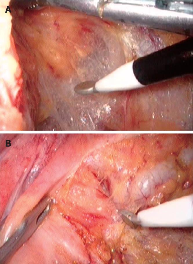
ES can produce a higher surface temperature when managing the tissues; the theoretical distance of thermal conduction is longer than that of US, which makes it easy to damage the surrounding tissues[31]. Therefore, it is particularly important for surgeons to understand how to use ES in laparoscopic TME with anal sphincter preservation for rectal cancer. The correct method is to maintain a certain tension on both sides of the separation space; let the ES slightly contact the tissue with its sharp edge, and then gently slide along the surface of tissue in order to form a large space (Figure 1A). The tissue was cut immediately into slide aside to expose deep layer tissue and the important organs in the deep layer tissue can easily be uncovered and avoid injury, thus reducing the possibility of the surrounding tissue necrosis due to “heat chemotactic effect”. At the important anatomical position, the operative speed must be slow, use “mm-class” dissection and sharp dissection in combination with blunt dissection in order to reduce bleeding and avoid injury of major blood vessels and organs (Figure 1B). There was no significant difference in postoperative complications such as anastomotic leakage and wound infection between ES group and US group in our study, indicating that ES is as safe as US in laparoscopic TME with anal sphincter preservation for rectal cancer. The operative time was almost the same between the two groups, although the cutting speed of ES was faster than that of US. On the other hand, the intraperitoneal CO2 must be exchanged regularly due to heavy smog generated by ES in order to maintain the clarity of operative vision, although it might slow the operative speed of ES in a certain extent. During operation, it must rely on the movement of metacarpophalangeal joint opening and closing repeatedly in order to control US. But in ES operation, surgeons might feel more comfortable as they can grasp ES, and rely on the push-pull movement of the arm, and the hand movement intensity might significantly decrease. Compared with US, ES has prominent advantages, including sturdiness, durability, low cost, and being suitable for laparoscopic TME with anal sphincter preservation for rectal cancer in less developed settings.

Figure 1
Figure 1 Intraoperative photograph. A: Showing monopolar ES dissect the space around the rectum; B: Showing monopolar ES dissect the seminal vesicle and the rectum.

In conclusion, ES is a safe and feasible tool similar to US used in laparoscopic TME with anal sphincter preservation for rectal cancer on the basis of skillful laparoscopic technique and complete understanding of laparoscopic pelvic anatomy. Application of ES can not only reduce the operation costs but also benefit the popularization of laparoscopic operation for rectal cancer patients.

COMMENTS
Background

Since the successful introduction of laparoscopic colectomy by Jacobs et al, laparoscopic surgery, especially laparoscopic rectal surgery has been developed considerably. Compared with open operation, the laparoscopic operation has many advantages such as less pain, little blood loss, small incision, good exposure of the pelvic cavity, an earlier return to daily activities, etc. while preserving the oncologic radicality of the procedure. However, the laparoscopic operation is more difficult than the open operation. Ultrasonically activated scalpel (US) is able to grasp and divide tissues while sealing small vessels, making the laparoscopic operation simpler, whereas the expensive medical instrument and high costs of the disposable materials can greatly increase the cost of laparoscopic rectal cancer operation.

Research frontiers

It has been shown that the laparoscopic rectal cancer operation had the same short-term and long-term outcomes compared with open surgery, so how to overcome the operation difficulty and reduce the cost of laparoscopic rectal operation has become hotspots in laparoscopic surgery.

Innovations and breakthroughs

In this study, authors tried to use cheap and durable monopolar electrocautery shovel (ES) in laparoscopic operation for rectal cancer in order to reduce the cost of the laparoscopic operation, they also summarized the technique and skill of using monopolar ES, and compared with US.

Applications

ES is a safe and feasible tool similar to US used in laparoscopic TME for rectal cancer on the basis of skillful laparoscopic technique and complete understanding of laparoscopic pelvic anatomy. Application of ES can not only reduce the operation costs but also facilitate the popularization of laparoscopic operation for rectal cancer.

Terminology

US is an expensive medical instrument which can incise tissue and seal small vessels, it is often used in complicated laparoscopic operations, but it costs high for the disposable materials. So in laparoscopic operation, US can greatly increase the cost of operation. ES is often used in simple laparoscopic operations because it is not as good as US for hemostasis, but it is cheap and durable.

Peer review

This is an interesting manuscript looking into two different methods of dissecting the rectum laparoscopically. It is a good paper.

Footnotes

Supported by Science and Technology Development Project of Hebei Province, No. 052761001D-26

References
1.  Jacobs M, Verdeja JC, Goldstein HS. Minimally invasive colon resection (laparoscopic colectomy). Surg Laparosc Endosc. 1991;1:144-150.  [PubMed]  [DOI]  [Cited in This Article: ]
2.  Staudacher C, Vignali A, Saverio DP, Elena O, Andrea T. Laparoscopic vs. open total mesorectal excision in unselected patients with rectal cancer: impact on early outcome. Dis Colon Rectum. 2007;50:1324-1331.  [PubMed]  [DOI]  [Cited in This Article: ]
3.  Liang JT, Lai HS, Lee PH, Huang KC. Comparison of functional and surgical outcomes of laparoscopic-assisted colonic J-pouch versus straight reconstruction after total mesorectal excision for lower rectal cancer. Ann Surg Oncol. 2007;14:1972-1979.  [PubMed]  [DOI]  [Cited in This Article: ]
4.  Pechlivanides G, Gouvas N, Tsiaoussis J, Tzortzinis A, Tzardi M, Moutafidis M, Dervenis C, Xynos E. Lymph node clearance after total mesorectal excision for rectal cancer: laparoscopic versus open approach. Dig Dis. 2007;25:94-99.  [PubMed]  [DOI]  [Cited in This Article: ]
5.  Lelong B, Bege T, Esterni B, Guiramand J, Turrini O, Moutardier V, Magnin V, Monges G, Pernoud N, Blache JL. Short-term outcome after laparoscopic or open restorative mesorectal excision for rectal cancer: a comparative cohort study. Dis Colon Rectum. 2007;50:176-183.  [PubMed]  [DOI]  [Cited in This Article: ]
6.  Uehara K, Yamamoto S, Fujita S, Akasu T, Moriya Y. Surgical outcomes of laparoscopic vs. open surgery for rectal carcinoma--a matched case-control study. Hepatogastroenterology. 2006;53:531-535.  [PubMed]  [DOI]  [Cited in This Article: ]
7.  Yano H, Ohnishi T, Kanoh T, Monden T. Hand-assisted laparoscopic low anterior resection for rectal carcinoma. J Laparoendosc Adv Surg Tech A. 2005;15:611-614.  [PubMed]  [DOI]  [Cited in This Article: ]
8.  Fukunaga M, Kidokoro A, Iba T, Sugiyama K, Fukunaga T, Nagakari K, Suda M, Yoshikawa S. Laparoscopy-assisted low anterior resection with a prolapsing technique for low rectal cancer. Surg Today. 2005;35:598-602.  [PubMed]  [DOI]  [Cited in This Article: ]
9.  Pigazzi A, Ellenhorn JD, Ballantyne GH, Paz IB. Robotic-assisted laparoscopic low anterior resection with total mesorectal excision for rectal cancer. Surg Endosc. 2006;20:1521-1525.  [PubMed]  [DOI]  [Cited in This Article: ]
10.  Ng SS, Lee JF, Yiu RY, Li JC, Hon SS. Telerobotic-assisted laparoscopic abdominoperineal resection for low rectal cancer: report of the first case in Hong Kong and China with an updated literature review. World J Gastroenterol. 2007;13:2514-2518.  [PubMed]  [DOI]  [Cited in This Article: ]
11.  Fukunaga Y, Higashino M, Tanimura S, Kishida S, Fujiwara Y, Ogata A, Osugi H. Laparoscopic mesorectal excision with preservation of the pelvic autonomic nerves for rectal cancer. Hepatogastroenterology. 2007;54:85-90.  [PubMed]  [DOI]  [Cited in This Article: ]
12.  Breukink SO, Pierie JP, Hoff C, Wiggers T, Meijerink WJ. Technique for laparoscopic autonomic nerve preserving total mesorectal excision. Int J Colorectal Dis. 2006;21:308-313.  [PubMed]  [DOI]  [Cited in This Article: ]
13.  Bretagnol F, Lelong B, Laurent C, Moutardier V, Rullier A, Monges G, Delpero JR, Rullier E. The oncological safety of laparoscopic total mesorectal excision with sphincter preservation for rectal carcinoma. Surg Endosc. 2005;19:892-896.  [PubMed]  [DOI]  [Cited in This Article: ]
14.  Tsang WW, Chung CC, Kwok SY, Li MK. Laparoscopic sphincter-preserving total mesorectal excision with colonic J-pouch reconstruction: five-year results. Ann Surg. 2006;243:353-358.  [PubMed]  [DOI]  [Cited in This Article: ]
15.  Gao F, Cao YF, Chen LS. Meta-analysis of short-term outcomes after laparoscopic resection for rectal cancer. Int J Colorectal Dis. 2006;21:652-656.  [PubMed]  [DOI]  [Cited in This Article: ]
16.  Dulucq JL, Wintringer P, Stabilini C, Mahajna A. Laparoscopic rectal resection with anal sphincter preservation for rectal cancer: long-term outcome. Surg Endosc. 2005;19:1468-1474.  [PubMed]  [DOI]  [Cited in This Article: ]
17.  Leroy J, Jamali F, Forbes L, Smith M, Rubino F, Mutter D, Marescaux J. Laparoscopic total mesorectal excision (TME) for rectal cancer surgery: long-term outcomes. Surg Endosc. 2004;18:281-289.  [PubMed]  [DOI]  [Cited in This Article: ]
18.  Liang JT, Lai HS, Lee PH, Huang KC. Comparison of functional and surgical outcomes of laparoscopic-assisted colonic J-pouch versus straight reconstruction after total mesorectal excision for lower rectal cancer. Ann Surg Oncol. 2007;14:1972-1979.  [PubMed]  [DOI]  [Cited in This Article: ]
19.  Asoglu O. A classical technique applied to laparoscopic rectal cancer surgery: transillumination of the inferior mesenteric root and its tributaries. J Laparoendosc Adv Surg Tech A. 2007;17:458-460.  [PubMed]  [DOI]  [Cited in This Article: ]
20.  Ishii Y, Hasegawa H, Nishibori H, Endo T, Kitajima M. The application of a new stapling device for open surgery (Contour Curved Cutter Stapler) in the laparoscopic resection of rectal cancer. Surg Endosc. 2006;20:1329-1331.  [PubMed]  [DOI]  [Cited in This Article: ]
21.  Kobayashi M, Morishita S, Okabayashi T, Miyatake K, Okamoto K, Namikawa T, Ogawa Y, Araki K. Preoperative assessment of vascular anatomy of inferior mesenteric artery by volume-rendered 3D-CT for laparoscopic lymph node dissection with left colic artery preservation in lower sigmoid and rectal cancer. World J Gastroenterol. 2006;12:553-555.  [PubMed]  [DOI]  [Cited in This Article: ]
22.  Fujii S, Shimada H, Ike H, Imada T, Yamagishi S, Saito S. Lifting of the colon for laparoscopic-assisted colectomy for colon and rectal cancer. JSLS. 2004;8:352-355.  [PubMed]  [DOI]  [Cited in This Article: ]
23.  Jitea N, Cristian D, Angelescu N. [Ultrasonically activated scalpel in laparoscopic surgery advantages and limits]. Chirurgia (Bucur). 2000;95:305-308.  [PubMed]  [DOI]  [Cited in This Article: ]
24.  Sheahan P, Miller I, Colreavy M, Sheahan JN, McShane D, Curran A. The ultrasonically activated scalpel versus bipolar diathermy for tonsillectomy: a prospective, randomized trial. Clin Otolaryngol Allied Sci. 2004;29:530-534.  [PubMed]  [DOI]  [Cited in This Article: ]
25.  Yamada S, Yoshino K, Inoue H. New-model ultrasonically activated shears for hemostatic sectioning during video-assisted thoracic surgery. Gen Thorac Cardiovasc Surg. 2007;55:518-520.  [PubMed]  [DOI]  [Cited in This Article: ]
26.  Nambirajan T, Woolsey S, Mahendra V, Walsh IK, Lynch TH, Keane PF. Efficacy and safety peri-prostatic local anaesthetic injection in trans-rectal biopsy of the prostrate: a prospective randomised study. Surgeon. 2004;2:221-224.  [PubMed]  [DOI]  [Cited in This Article: ]
27.  Amaral JF. The experimental development of an ultrasonically activated scalpel for laparoscopic use. Surg Laparosc Endosc. 1994;4:92-99.  [PubMed]  [DOI]  [Cited in This Article: ]
28.  Foschi D, Cellerino P, Corsi F, Taidelli T, Morandi E, Rizzi A, Trabucchi E. The mechanisms of blood vessel closure in humans by the application of ultrasonic energy. Surg Endosc. 2002;16:814-819.  [PubMed]  [DOI]  [Cited in This Article: ]
29.  Wara P, Berg V, Jacobsen NO, Casalnuovo C, Amdrup E. Possible mechanism of hemostasis effected by electrocoagulation. Endoscopy. 1984;16:43-46.  [PubMed]  [DOI]  [Cited in This Article: ]
30.  Morris ML. Electrosurgery in the gastroenterology suite: principles, practice, and safety. Gastroenterol Nurs. 2006;29:126-132; quiz 132-134.  [PubMed]  [DOI]  [Cited in This Article: ]
31.  Antonutti R, Fontes-Dislaire I, Rumeau JL, Mutter D, Fourtanier G. [Experimental study of monopolar electrical and ultrasonic dissection]. Ann Chir. 2001;126:330-335.  [PubMed]  [DOI]  [Cited in This Article: ]