Copyright
©2007 Baishideng Publishing Group Inc.
World J Gastroenterol. Nov 21, 2007; 13(43): 5787-5793
Published online Nov 21, 2007. doi: 10.3748/wjg.v13.i43.5787
Published online Nov 21, 2007. doi: 10.3748/wjg.v13.i43.5787
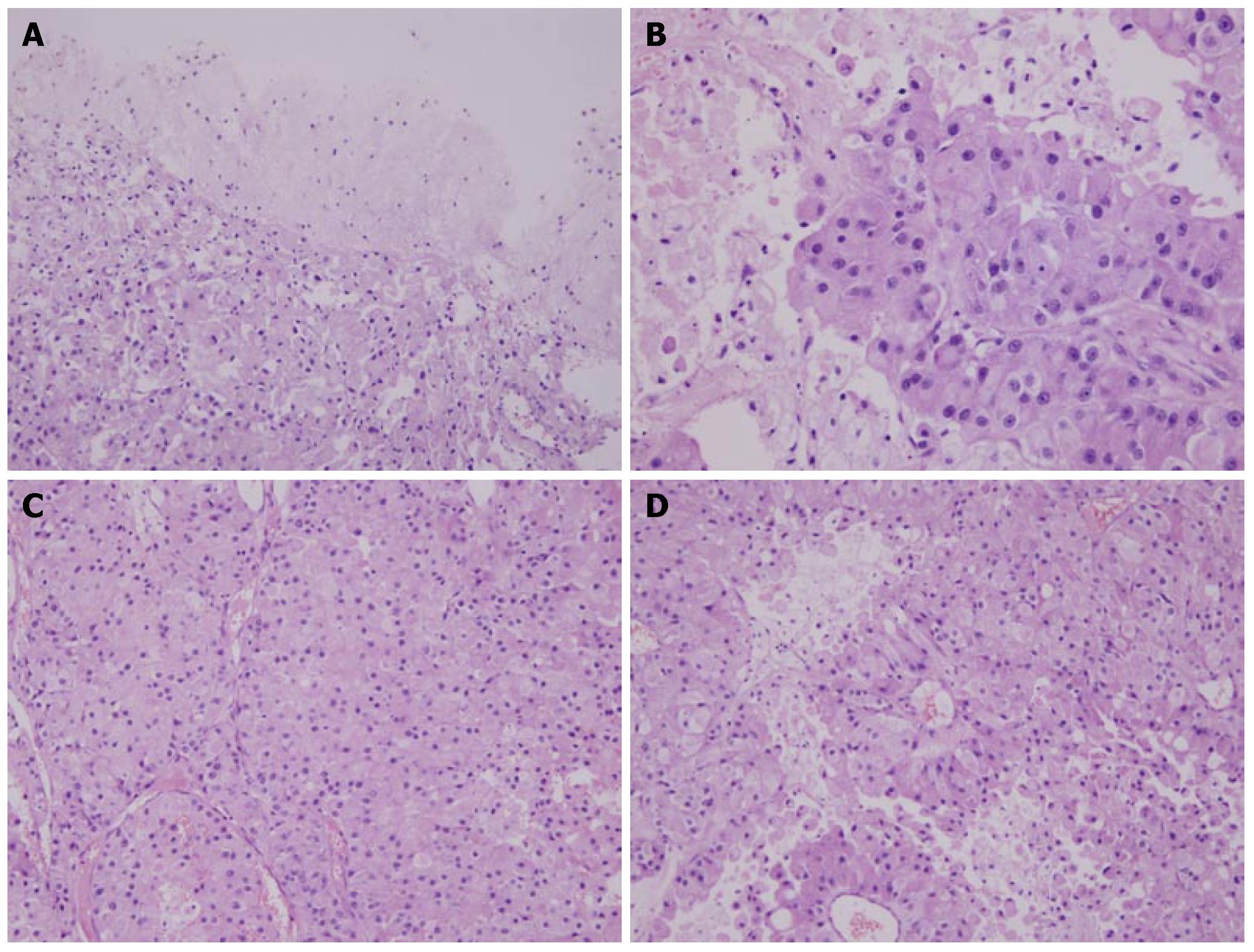
Figure 2 Brain metastatic tumor showing the growth pattern of solid cell nests (HE stain).
A: Polygonal tumor cells with abundant eosinophilic cytoplasm, rich blood vessels and clear boundary of tumor and brain parenchyma (× 100); B: Polygonal tumor cells showing epitheliod and abundant eosinophilic cytoplasm, rich chromatin nuclei with obvious nucleoli (× 200); C: Round nuclei with obvious nucleoli, rich blood vessels resembling sinusoid-like blood spaces in hepatocellular carcinoma (× 200); D: Tumor cells exhibiting radial pattern surrounding thin- walled vessels (× 200).
- Citation: Zhang S, Wang M, Xue YH, Chen YP. Cerebral metastasis from hepatoid adenocarcinoma of the stomach. World J Gastroenterol 2007; 13(43): 5787-5793
- URL: https://www.wjgnet.com/1007-9327/full/v13/i43/5787.htm
- DOI: https://dx.doi.org/10.3748/wjg.v13.i43.5787


