Copyright
©2010 Baishideng.
World J Gastroenterol. Mar 7, 2010; 16(9): 1076-1085
Published online Mar 7, 2010. doi: 10.3748/wjg.v16.i9.1076
Published online Mar 7, 2010. doi: 10.3748/wjg.v16.i9.1076
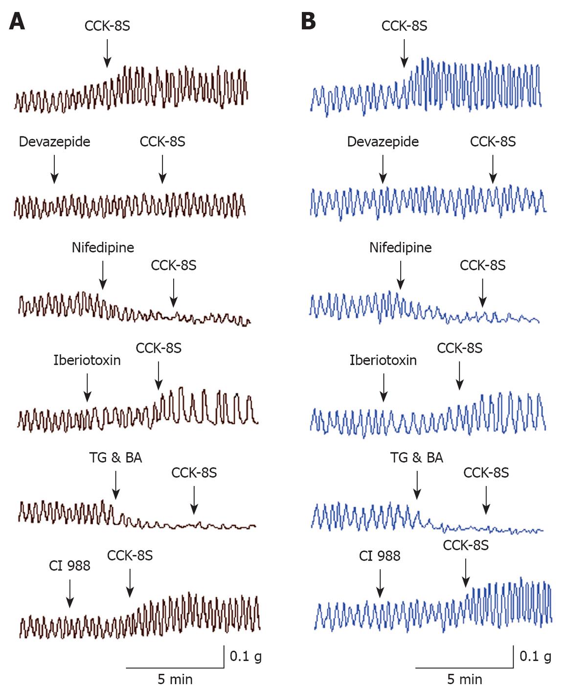
Figure 1 Effects of CCK-8S on the resting tension of guinea pig proximal colonic smooth muscle (PCSM) strips.
A: Representative traces of the effect of CCK-8S (10-7 mol/L) on the resting tension of circular muscle (CM) strips. CCK-8S increased the mean contractile amplitude of CM strips, which was inhibited by pretreatment with CCK1 receptor antagonist devazepide (10-7 mol/L), L-type calcium channel inhibitor nifedipine (10-5 mol/L), Ca2+-ATPase inhibitor thapsigargin (TG) (10-5 mol/L) and intracellular calcium chelator BAPTA-AM (BA) (10-5 mol/L). Pretreating CM strips with iberiotoxin (10-6 mol/L), a selective potassium channel inhibitor, did not block the CCK-8S-induced increase in the contractile amplitude of CM strips but decreased the frequency; B: Representative traces of the effect of CCK-8S (10-7 mol/L) on the resting tension of longitudinal muscle (LM) strips. The CCK-8S-induced increase in the amplitude and frequency of LM strips was inhibited by pretreatment with devazepide (10-7 mol/L), nifedipine (10-5 mol/L) or TG (10-5 mol/L) and BA (10-5 mol/L). Pretreating LM strips with iberiotoxin (10-6 mol/L) did not attenuate the CCK-8S-induced increase in the contractile amplitude of LM strips but decreased the frequency. CCK2 receptor antagonist CI 988 had no effect on the CCK-8S-induced contraction of CM and LM strips.
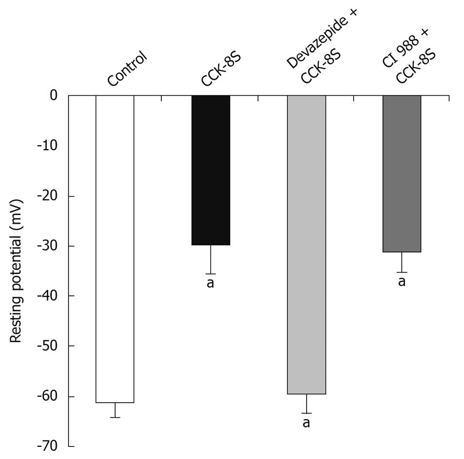
Figure 2 Effects of CCK-8S on resting potential (RP) of PCSMCs.
RP rose from -61.3 ± 2.7 mV to -29.8 ± 5.9 mV after superfusion with CCK-8S (10-7 mol/L). In the presence of devazepide (10-7 mol/L), CCK-8S (10-7 mol/L) did not depolarize RP (-59.4 ± 3.8 mV). When SMCs were pretreated with CI 988, CCK-8S (10-7 mol/L) depolarized RP to -29.8 ± 5.9 mV. aP < 0.01 vs control group (CaPSS).
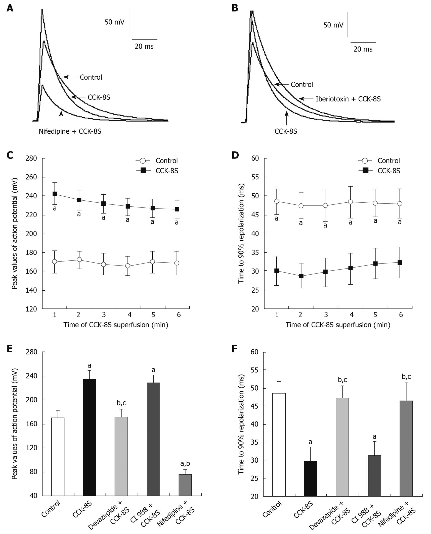
Figure 3 Effects of CCK-8S on AP of PCSMCs.
A-C and E: CCK-8S (10-7 mol/L) suppressed the amplitude of AP to 38.6% ± 3.2% (n = 15, P < 0.01); A, B, D and F: CCK-8S (10-7 mol/L) decreased fast repolarization times (T90) by 36.9% ± 8.7% (n = 15, P < 0.01); A and E: Nifedipine (10-5 mol/L) blocked the effects of CCK-8S on the amplitude of AP but failed to have any effect on T90; B and F: Iberiotoxin (10-6 mol/L) blocked the effects of CCK-8S on T90 but failed to have any effect on the amplitude of AP; E and F: Pretreating SMCs with devazepide (10-7 mol/L) blocked the CCK-8S-evoked effect on AP amplitude and T90, whereas CI 988 had no such effect. aP < 0.01 vs control group (CaPSS); bP < 0.01 vs CCK-8S group; cP > 0.05 vs control group.
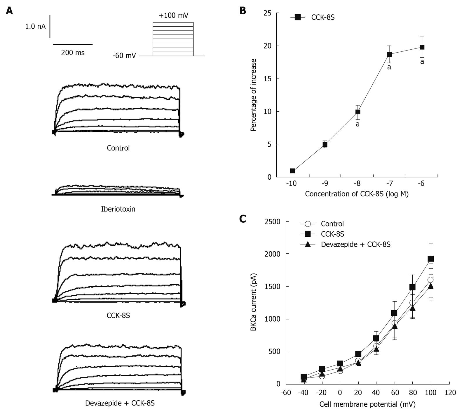
Figure 4 Effects of CCK-8S on IBKCa of PCSMCs.
A: Current traces of IBKCa were recorded from -60 mV to +100 mV during 400 ms depolarization; B: CCK-8S increased IBKCa in a concentration-dependent manner (EC50 = 3.5 × 10⁻⁸ mol/L); CCK-8S (10-7 mol/L) enhanced IBKCa depolarized from -60 mV to +60 mV (from 916 ± 183 pA to 1088 ± 226 pA; n = 8, P < 0.01); C: Current-voltage relationship of average peak IBKCa under each condition. CCK-8S-evoked enhancement of IBKCa was suppressed by devazepide (10-7 mol/L). aP < 0.05 vs control group (CaPSS).
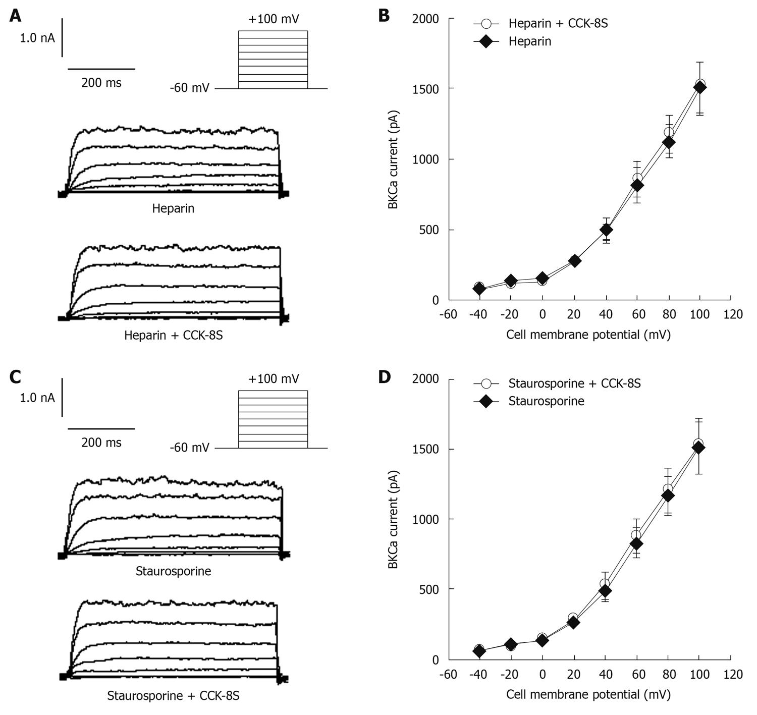
Figure 5 Heparin and staurosporine prevented CCK-8S-intensified IBKCa in PCSMCs.
A and C: Current traces of IBKCa were evoked by a step from -60 mV to +60 mV; B: When heparin (10-6 mol/L) was present in the pipette solution, CCK-8S (10-7 mol/L) did not increase IBKCa; D: CCK-8S (10-7 mol/L) failed to increase IBKCa in the presence of staurosporine (10-6 mol/L).
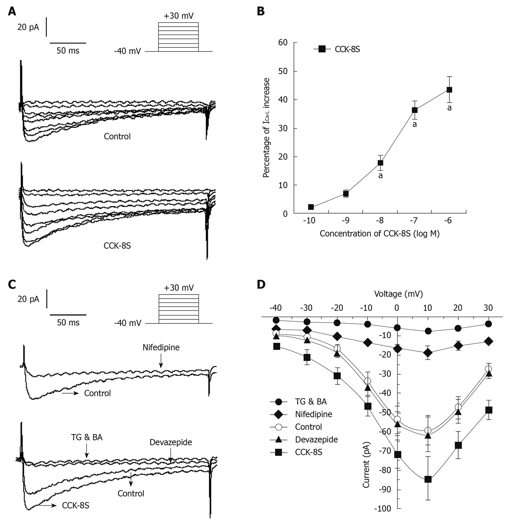
Figure 6 Effects of CCK-8S, nifedipine and devazepide on ICa-L in PCSMCs.
A: Current traces of ICa-L were recorded from -40 mV to +30 mV during 400 ms depolarization; B: CCK-8S enhanced ICa-L in a concentration-dependent manner (EC50 = 3.2 × 10⁻⁸ mol/L); CCK-8S (10-7 mol/L) increased ICa-L depolarized from -40 mV to +10 mV (from 60 ± 8 pA to 84 ± 11 pA; n = 8); C: CCK-8S-intensified ICa-L was markedly suppressed by devazepide (10-7 mol/L), nifedipine (10-5 mol/L), TG and BA (10-5 mol/L); D: Current-voltage curves of whole-cell patch clamp under each condition. Compared with control, CCK-8S (10-7 mol/L) significantly enhanced ICa-L, whereas devazepide, nifedipine, TG and BA inhibited ICa-L. aP < 0.05 vs control group (CaPSS).
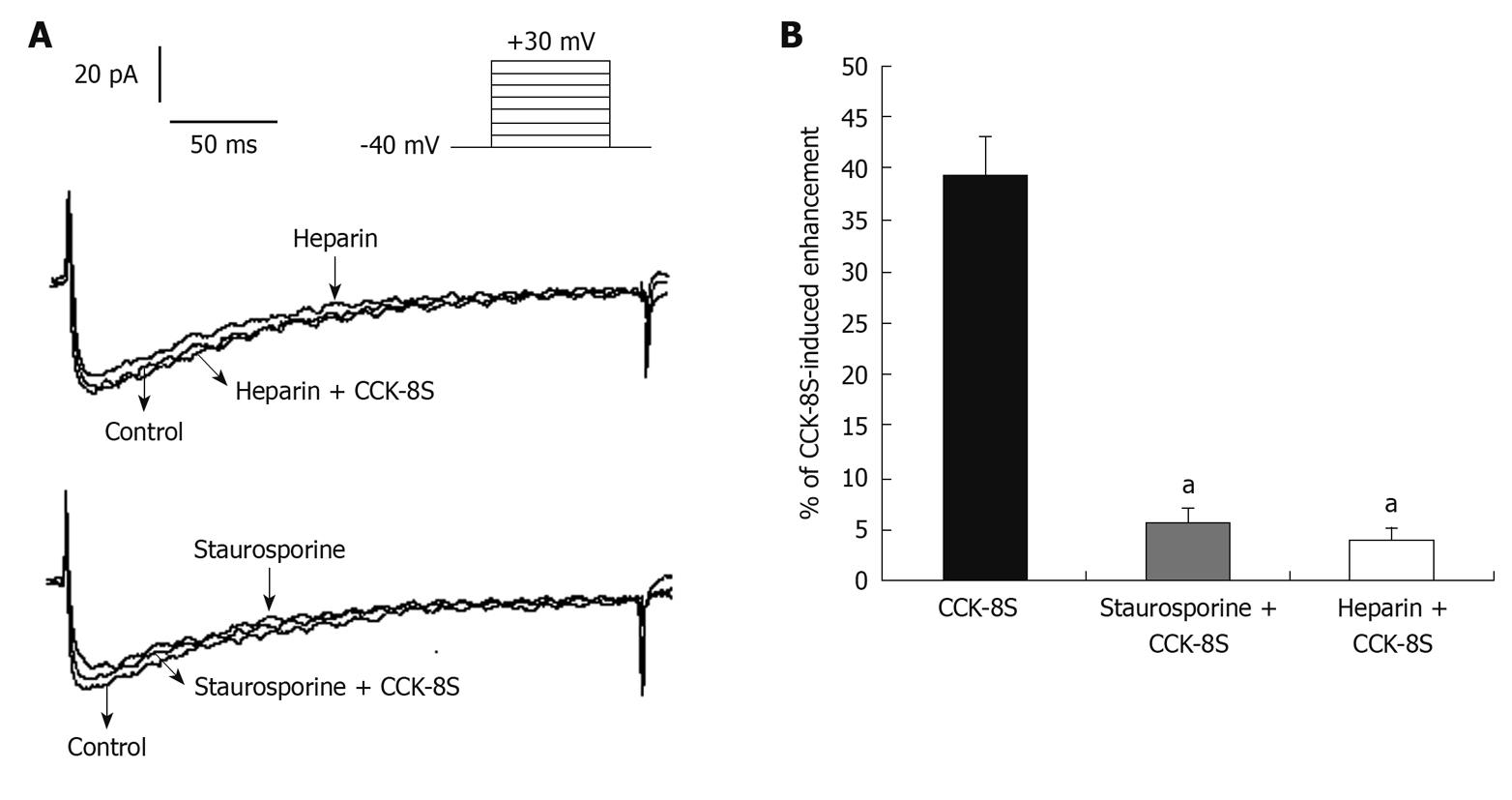
Figure 7 Heparin and staurosporine prevented CCK-8S-intensified ICa-L in PCSMCs.
A: Current traces of ICa-L were evoked by a step from -40 mV to +10 mV. When heparin (10-6 mol/L) was present in the pipette solution, CCK-8S (10-7 mol/L) did not increase IBKCa; CCK-8S (10-7 mol/L) failed to increase IBKCa in the presence of staurosporine (10-6 mol/L); B: CCK-8S-induced enhancement of ICa-L in the absence and presence of staurosporine and heparin, respectively. aP < 0.05 vs CCK-8S group.
- Citation: Zhu J, Chen L, Xia H, Luo HS. Mechanisms mediating CCK-8S-induced contraction of proximal colon in guinea pigs. World J Gastroenterol 2010; 16(9): 1076-1085
- URL: https://www.wjgnet.com/1007-9327/full/v16/i9/1076.htm
- DOI: https://dx.doi.org/10.3748/wjg.v16.i9.1076


