Copyright
©2010 Baishideng.
World J Gastroenterol. Feb 21, 2010; 16(7): 909-913
Published online Feb 21, 2010. doi: 10.3748/wjg.v16.i7.909
Published online Feb 21, 2010. doi: 10.3748/wjg.v16.i7.909
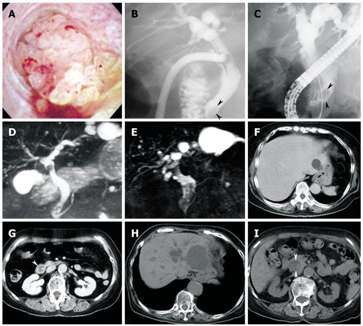
Figure 1 Images of cholangioscopic examination, ERC, MRCP and abdominal CT.
A: Cholangioscopic examination performed in 1999. A multilobulated papillary tumor was seen protruding into the common bile duct; B: ERC performed in 1999; C: ERC performed in 2008. The extrahepatic bile duct became more dilated in 2008 than in 1999. A polypoid filling defect (arrow heads) could be detected in the distal bile duct; D: MRCP in 1999; E: MRCP in 2004. Cystic lesions connected to the intrahepatic bile ducts of the left liver became enlarged in 2004; F and G: Abdominal CT in 1999; H and I: Abdominal CT in 2004. Intra- and extrahepatic bile ducts became dilated in 2004. A mass in the distal bile duct (arrow heads) and a cystic lesion in the left liver were enlarged in 2004, compared with those in 1999.
Figure 2 Images of ultrasonographic studies.
A: EUS showed a mixed echoic mass in the distal bile duct (arrow heads); B: Papillary structure of the mass could be apparently observed on CEH-EUS with Sonazoid®; C: IDUS showed a 10-mm papillary mass (arrows) in the middle bile duct; D: In the distal bile duct, multilobular and papillary lesions (arrows) were observed. L: Lumen of the bile duct.
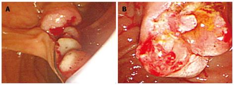
Figure 3 EST was performed.
A: A soft, bead-like mass was extracted through the orifice of the ampulla of Vater after EST; B: Image of the ampulla of Vater after extraction of the mass by balloon sweep and a net-type catheter.
Figure 4 Histopathological findings.
A: Hematoxylin-eosin staining; B: Immunohistochemical analysis for MUC1; C: Immunohistochemical analysis for MUC5AC.
- Citation: Tsuchida K, Yamagata M, Saifuku Y, Ichikawa D, Kanke K, Murohisa T, Tamano M, Iijima M, Nemoto Y, Shimoda W, Komori T, Fukui H, Ichikawa K, Sugaya H, Miyachi K, Fujimori T, Hiraishi H. Successful endoscopic procedures for intraductal papillary neoplasm of the bile duct: A case report. World J Gastroenterol 2010; 16(7): 909-913
- URL: https://www.wjgnet.com/1007-9327/full/v16/i7/909.htm
- DOI: https://dx.doi.org/10.3748/wjg.v16.i7.909


