Published online Aug 24, 2017. doi: 10.5500/wjt.v7.i4.222
Peer-review started: February 8, 2017
First decision: May 8, 2017
Revised: June 6, 2017
Accepted: June 30, 2017
Article in press: July 3, 2017
Published online: August 24, 2017
To compare the differential immune T cell subset composition in patients with acute T cell-mediated rejection in the kidney transplant with subset composition in the absence of rejection, and to explore the association of their respective immune profiles with kidney transplant outcomes.
A pilot cross-sectional histopathological analysis of the immune infiltrate was performed using immunohistochemistry in a cohort of 14 patients with acute T cell-mediated rejection in the kidney transplant and 7 kidney transplant patients with no rejection subjected to biopsy to investigate acute kidney transplant dysfunction. All patients were recruited consecutively from 2012 to 2014 at the Singapore General Hospital. Association of the immune infiltrates with kidney transplant outcomes at up to 54 mo of follow up was also explored prospectively.
In comparison to the absence of rejection, acute T cell-mediated rejection in the kidney transplant was characterised by numerical dominance of cytotoxic T lymphocytes over Foxp3+ regulatory T cells, but did not reach statistical significance owing to the small sample size in our pilot study. There was no obvious difference in absolute numbers of infiltrating cytotoxic T lymphocytes, Foxp3+ regulatory T cells and Th17 cells between the two patient groups when quantified separately. Our exploratory analysis on associations of T cell subset quantifications with kidney transplant outcomes revealed that the degree of Th17 cell infiltration was significantly associated with shorter time to doubling of creatinine and shorter time to transplant loss.
Although this was a small pilot study, results support our suspicion that in kidney transplant patients the immune balance in acute T cell-mediated rejection is tilted towards the pro-rejection forces and prompt larger and more sophisticated studies.
Core tip: In the clinical setting, acute T cell-mediated rejection in the kidney transplant (ATCMR-KTx) is only confirmed through a kidney transplant biopsy, which is scored according to the Banff classification. The Banff classification is largely based on the estimation of mononuclear cell infiltration instead of the identification and quantification of the actual T cell subsets recruited to mediate rejection. Therefore, a more detailed analysis of the inflammatory infiltrate of ATCMR-KTx is likely to enhance the diagnostic accuracy of the Banff classification. In our analyses, ATCMR-KTx appeared to be characterised by a numerical dominance of cytotoxic T lymphocytes over regulatory T cells in comparison to the absence of acute rejection. We also found an association of the numbers of infiltrating Th17 cells with kidney transplant outcomes. Although this is a small pilot study, it further supports our suspicion that the immune balance in ATCMR-KTx is tilted towards the pro-rejection forces.
- Citation: Salcido-Ochoa F, Hue SSS, Peng S, Fan Z, Li RL, Iqbal J, Allen Jr JC, Loh AHL. Histopathological analysis of infiltrating T cell subsets in acute T cell-mediated rejection in the kidney transplant. World J Transplant 2017; 7(4): 222-234
- URL: https://www.wjgnet.com/2220-3230/full/v7/i4/222.htm
- DOI: https://dx.doi.org/10.5500/wjt.v7.i4.222
Acute T cell-mediated rejection in the kidney transplant (ATCMR-KTx) is a common encounter in kidney transplantation. It can perpetuate itself as chronic T cell-mediated rejection or transform into antibody-mediated rejection, which progressively can destroy the renal parenchyma, leading to reduction of kidney transplant survival with potential transplant loss and the return to dialysis[1,2]. Therefore, adequate maintenance immunosuppression to prevent the occurrence of ATCMR-KTx, prompt and accurate identification, and early initiation of anti-rejection therapy are needed to minimise patient’s complications and to improve long-term kidney transplant outcomes.
In the current state of the art, confirmation of ATCMR-KTx is based on scoring kidney transplant histopathological changes using the Banff classification[3]. Despite being the gold standard, there are a few limitations. The Banff classification relies on a semi-quantitative estimation of the infiltrating mononuclear cells. This approach, however, does not distinguish the actual cellular program that is operating within the transplant tissue. We believe that identification of the actual T cell subsets infiltrating the kidney transplant provides better insight into the immunologic events in ATMCR-KTx. In other words, a more detailed analysis of the inflammatory infiltrate of kidney transplant biopsies undergoing ATCMR-KTx is expected to reflect more accurately the status of alloactivation within the kidney transplant and to lead to a better understanding of the immunopathogenesis of ATCMR-KTx. Similarly, this information could be used in the future to improve the accuracy and the predictive value of the Banff classification in kidney transplantation.
The immunologically-mediated damage of ATCMR-KTx is mediated and executed by different subtypes of effector T cells, including cytotoxic T lymphocytes (CTL), T helper (Th) 17 cells and Th1 cells, as well as natural killer cells and monocytes. In addition, Foxp3+ regulatory T cells (Treg cells) are also known to migrate to the transplant tissue to modulate the inflammatory response[4-9].
CTL are central effectors of alloimmune damage to the parenchymal cells of the kidney transplant[10,11]. Therefore, the detection of their cytotoxic products inside the kidney transplant is commonly used as a surrogate of their presence and their allotoxicity. To highlight a few examples, at the molecular level intra-graft detection of granzyme B mRNA has been shown to be able to differentiate ATCMR-KTx from the absence of rejection[12,13]. Concomitant detection of both granzyme B and perforin mRNA[14,15], or of both granzyme B and CD178 mRNA[13,16] have also been shown to identify ATCMR-KTx with higher accuracy. It has also been reported that the detection of granulysin mRNA, another CTL product, helped to differentiate patients with ATCMR-KTx from those with no rejection in their biopsies[17]. A similar result has also been observed at the protein level by immunohistochemical detection of granzyme B and perforin expression[10]. Although the outcome of kidney transplantation after an episode of ATCMR-KTx is difficult to predict, there are some indications that the detection of markers of CTL in the kidney transplant may offer some value. One study demonstrated that a higher degree of granzyme B+ cell infiltration in the allograft was associated with poorer allograft survival[18], and another study showed that the intra-graft expression of granzyme B was associated with the severity of the rejection process[10]. Likewise, the expression of CD178[19] or the co-expression of both CD178 and granzyme B[13] conferred poorer prognosis to patients suffering from ATCMR-KTx. Despite the aforementioned findings, it has been suggested that expression of granzyme B by itself may have limited clinical predictive value[19].
Th17 cells are another type of effector T cells involved in alloimmunity and in biopsies are usually identified by the detection of IL-17. It has been reported that the magnitude of Th17 cell infiltration over Treg cell infiltration correlated with kidney transplant function, the degree of interstitial inflammation and tubular atrophy, the refractoriness to treatment and the recurrence of ATCMR-KTx[20-22].
Despite the belief that Th1 cells are believed to be crucial mediators of the rejection process, the detection of interferon-gamma, as a surrogate marker for their presence, was no better than the detection of cytotoxic molecules for the diagnosis of pure ATCMR-KTx[13]. In addition, intra-graft expression of T-bet, also a surrogate marker for Th1 cells, was not able to distinguish ATCMR-KTx from the absence of rejection. In this respect, the role of Th2 cells in the rejection process appears to be less dramatic and less understood; and the identification of Th2 cells through the detection of intra-graft IL-4 mRNA was also not useful for the diagnosis of ACTMR-KTx[13].
Although several reports have implicated Foxp3+ Treg cells in alloregulation and transplantation tolerance in animal models[8,23] and in humans[24], the detection of Foxp3+ Treg cells to aid in the diagnosis of ATCMR in the kidney transplant and their clinical significance has been beset with controversy[25]. Some authors have published that higher infiltration by Foxp3+ Treg cells appeared to associate with more favourable transplant outcomes in patients with ATCMR-KTx[26] and in patients with subclinical rejection found in protocol biopsies[27,28], in comparison to those cases of much lower infiltration by Foxp3+ Treg cells. Likewise, patients with ATCMR-KTx having higher expression of Foxp3 mRNA were more likely to respond to therapy that those with lower levels[20]. However, other studies reported were not very supportive of the detection of Treg cells in ATCMR-KTx. The detection of intra-graft Foxp3 mRNA, as a surrogate marker for Foxp3+ Treg cells, was not associated with the diagnosis ATCMR-KTx in one study[12]. In addition, no association was found in another study of ATCMR-KTx between the detection of Foxp3+ T cells by immunofluorescence and kidney transplant outcomes[29].
We have hypothesised that the balance between effector and Foxp3+ Treg cells could play a role in determining the occurrence and severity of ATCMR-KTx, as well as predicting the potential outcome of the kidney transplant[25]. However, as discussed previously, the clinical significance of the immune infiltrate in ATCMR-KTx or its balance is controversial. Therefore, in this study performed in a cohort of Asian patients, we aimed to identify and quantify the main T cell subsets infiltrating the kidney transplant undergoing ATCMR and to compare with that in the absence of rejection. We use immunohistochemistry as our detection technology as it is inexpensive, easily reproducible and accessible to many laboratories. Based on the literature presented above, we focused our immunodetection on the most promising markers, i.e., granzyme B and IL-17 (representing CTL and Th17 cells, respectively) and Foxp3 (representing Foxp3+ Treg cells). To assess their immune balance, we arbitrarily measured their numerical ratios within the immune infiltrate found in both kidney transplant patients with ATCMR-KTx and with no rejection. Then, we explored the association of the numbers of these subsets and their ratios with kidney transplant outcomes up to fifty-four months of clinical follow up. We focused our outcome analysis on the risk of subsequent rejection episodes, deterioration of kidney transplant function and immunologically-mediated transplant loss.
Cross-sectional immunohistochemical analysis performed in formalin-fixed paraffin-embedded tissue collected in a consecutive cohort of 21 kidney transplant patients that were subjected to kidney transplant biopsy for the investigation of acute kidney transplant dysfunction at any time post-transplantation. Patients satisfying our inclusion and exclusion criteria were subdivided post-hoc into two groups: (1) ATCMR-KTx; and (2) no rejection. All patients were recruited between 1 January 2012 to 1 January 2014 at the Singapore General Hospital (SGH), the largest tertiary care and academic centre in Singapore; and followed for kidney transplant outcomes up to fifty-four months from the time of transplant biopsy.
Inclusion criteria: Adult kidney transplant patients (aged 21-80 years) who were of low immunological risk (ABO-compatible, lack of donor-specific antibodies, negative cross-match, no history of antibody-mediated rejection); who had acute kidney transplant dysfunction due to: (1) ATCMR-KTx (category 4 of the Banff 2009 classification); or (2) found with absence of rejection in the biopsy (category 1 of the Banff 2009 classification, or category 6 of the Banff 2009 classification of no inflammatory or infective nature, i.e., with no BK virus nephropathy, other infections affecting the transplant, glomerulonephritis or interstitial nephritis).
Exclusion criteria: Human immunodeficiency virus infection, history of haematological malignancies, children, pregnant women, poor cognitive capacity, prisoners and the inability to understand the research protocol and give consent. Patients whose biopsies showed borderline rejection (category 3 of the Banff 2009 classification) or antibody-mediated rejection (category 2 of the Banff 2009 classification) were also excluded from the analysis. Biopsies in the non-rejection group were revised according to the Banff 2013 update before the final analysis, to ensure they still satisfy the non-rejection group criteria according to the Banff 2013 update.
Baseline demographic and clinical characteristics as well as clinical outcomes were retrieved from clinical hard-copy case notes and our electronic medical records. Use and type of immunosuppressants prescribed were also recorded.
Serum creatinine and urine protein to creatinine ratio (or total urinary protein in a 24-h collection) were measured. Calculated estimated glomerular filtration rate (eGFR) was obtained through the “modification of diet in renal disease” equation. All laboratory parameters were retrieved prospectively from electronic medical records from the time of kidney transplant biopsy and at 3, 6, 12, 18, 24, 30, 36, 42, 48 and 54 mo of follow up post-biopsy. All laboratory investigations were conducted at the SGH’s clinical laboratory, which is accredited by the College of American Pathologists.
Immunohistochemistry for detection of T cell subsets in kidney transplant tissue biopsies was performed in both the Renal Laboratory and the Pathology Laboratory at the SGH. In brief, slides prepared from formalin-fixed paraffin-embedded kidney tissue specimens were stained with monoclonal antibodies conjugated with either horseradish peroxidase or alkaline phosphatase and directed against different phenotypic markers, including CD4, CD8, CD19, IL-17, granzyme B and Foxp3. The binding of the different antibodies onto the kidney tissue samples was revealed using the respective chromogenic substrates for those enzymes. Isotype-matched antibodies were used as negative controls. Tonsil tissue served as positive control. Staining was visualized and quantified directly by light microscopy and adjusted to biopsy tubulo-interstitial area (vessels and glomeruli excluded) measured by Olympus CellSens software. Percentage of infiltration of CD4+, CD8+ and CD19+ cells, as well as the number of Foxp3-, IL-17- or granzyme B-expressing cells per square millimetre of kidney tubulo-interstitial area in the biopsy (cell density) was reported. The ratios between the cell densities of granzyme B- and IL-17-expressing cells over Foxp3-expressing cells were calculated.
Sample size: As this was an exploratory study on consecutively recruited patients, sample size was not calculated a priori.
To determine whether tissue-infiltrating T cell profiles differ between kidney transplant patients with: (1) biopsy-proven ATCMR-KTx; and (2) no rejection, median cell densities of tissue-infiltrating: (1) granzyme B+ CTL; (2) IL-17+ Th17 cells; (3) Foxp3+ Treg cells were compared between these two groups of patients. In addition, ratios of the cell densities of tissue-infiltrating; (4) granzyme B+ CTL over Foxp3+ Treg cells; and of (5) IL-17+ Th17 cells over Foxp3+ Treg cells were compared between kidney transplant patients with: (1) biopsy-proven ATCMR-KTx; and (2) no rejection. Medians were compared using the Wilcoxon rank-sum test. Spearman correlation was used to assess strength of association of densities and ratios of infiltrating immune cells with different kidney transplant outcomes, including: (1) changes in serum creatinine; (2) eGFR; and (3) proteinuria. Longitudinal analysis of variance was used to display and compare changes in these same outcome variables between the two groups of patients over the follow up period. The analysis was performed on log-transformed values in order to achieve normality of residuals. The log-rank test was used to compare time-to-event curves between the biopsy-proven ATCMR-KTx and the no-rejection groups for the following outcomes: (1) time to any rejection (a composite outcome including borderline rejection, ATCMR-KTx or antibody-mediated rejection occurring post-biopsy during the follow up period); (2) time to doubling of creatinine post-biopsy; and (3) time to confirmed or suspected immune-mediated transplant loss. The date for re-initiation of dialysis was taken as the date of transplant loss. Cox regression analysis was used to investigate the effect of the cell densities of the tissue infiltrating T cells and their ratios and other clinical parameters (potential confounders taken from Table 1) on different kidney transplant outcomes including: (1) time to any rejection post-biopsy; (2) time to doubling of creatinine post-biopsy; and (3) time to confirmed or suspected immune-mediated transplant loss. All analyses were performed using SAS V9.4 software (SAS Inc., Cary NC, United States).
| Characteristic | n3 | No rejection | n4 | ATCMR | P value |
| Age (yr)1 | 7 | 60.8 | 14 | 44.9 | 0.0101 |
| Male sex (%) | 7 | 57.14 | 14 | 71.43 | 0.6384 |
| Race Chinese (%) | 7 | 86.71 | 14 | 57.14 | 0.3371 |
| Dialysis vintage (yr)1 | 7 | 2.08 | 14 | 5.015 | 0.6888 |
| Transplant vintage (yr)1 | 7 | 13.75 | 14 | 3.935 | 0.0031 |
| Deceased donor (%) | 6 | 66.67 | 13 | 53.85 | > 0.9999 |
| Delayed graft function (%) | 6 | 33.33 | 12 | 41.67 | > 0.9999 |
| Cold ischaemia time (h) | 5 | 3 | 9 | 10 | 0.6973 |
| Total HLA mismatch (#)1 | 6 | 3 | 11 | 3 | 0.9973 |
| Very high immune risk (%)2 | 6 | 16.67 | 11 | 43.45 | 0.3334 |
| % Panel of reactive antibodies1 | 3 | 8 | 9 | 0 | 0.2318 |
| History of ATCMR (%) | 7 | 14.29 | 14 | 50 | 0.1736 |
| Re-transplant (%) | 7 | 0 | 14 | 7.14 | > 0.9999 |
| GFR at biopsy (mL/min per 1.73 m2)1 | 7 | 41.2 | 14 | 17.95 | 0.0767 |
| Proteinuria at biopsy (g/d)1 | 7 | 3.5 | 14 | 1.23 | 0.2028 |
| t score1 | 7 | 0 | 14 | 2 | 0.0116 |
| I score1 | 7 | 1 | 14 | 2 | 0.0007 |
| v score1 | 7 | 0 | 14 | 0 | 0.1196 |
| Tacrolimus use at biopsy (%) | 7 | 0 | 14 | 50 | 0.0468 |
| Ciclosporin use at biopsy (%) | 7 | 100 | 14 | 35.71 | 0.0071 |
| MTORI use at biopsy (%) | 7 | 0 | 14 | 14.29 | 0.5333 |
| Steroids use at biopsy (%) | 7 | 100 | 14 | 100 | > 0.9999 |
| Mycophenolate use at biopsy (%) | 7 | 57.14 | 14 | 85.71 | 0.28 |
| Azathioprine use at biopsy (%) | 7 | 28.57 | 14 | 0 | 0.10 |
| Anti-CD25 induction (%) | 5 | 0 | 12 | 41.67 | 0.2445 |
| Prior thymoglobulin use (%) | 7 | 14.29 | 14 | 14.29 | > 0.9999 |
Table 1 shows the main clinical and demographic characteristics of the 21 recruited kidney transplant patients that were subjected to transplant biopsy for the investigation of acute kidney transplant dysfunction (14 with ATCMR-KTx and 7 with no rejection, inflammation nor infection found in their biopsy). Overall, in the ATCMR-KTx group the need for transplant biopsy occurred earlier post-transplantation than for the non-rejection group and had worse kidney function at presentation. History of previous rejection episodes occurred preferentially in this group too. They also had slightly higher rate of delayed graft function and longer cold-ischaemia than the non-rejection group. The HLA mismatches and the immune risk according to Fuggle’s classification[30] was similar in both groups. In our patient cohort, all the non-rejection patients were taking ciclosporin as maintenance immunosuppression at the time of the biopsy, while half of the patients in the ATCMR-KTx group were on tacrolimus. The acute rejection scores (t, i and v) of the Banff classification were higher in the ATCMR-Tx group, as expected. Tables 2 and 3 provide the detailed clinical and demographic characteristics of each recruited patient, as well as their particular immune variables and main kidney transplant outcomes.
| Patient | Group | Age | Sex | Race | Dialysis vintage (yr) | Tx vintage (yr) | Donor type | DGF | CIT (h) | HLA MM (#) | Immune risk | PRA (%) | ATCMR Hx | Re-Tx | Anti-CD25 induction | ATG use | Immuno-suppression at Bx |
| 1 | ATCMR | 49.9 | M | Ma | 0.36 | 14.26 | Living | No | 0 | 0 | Low | UNK | Yes | No | No | No | CsA + MPA |
| 2 | ATCMR | 32.1 | F | Ch | 0.38 | 0.17 | Living | No | UNK | 1 | Very high | 20 | Yes | No | Yes | No | MTORI + MPA |
| 3 | ATCMR | 25.7 | M | Ma | 1.21 | 6.80 | UNK | UNK | UNK | UNK | UNK | UNK | Yes | No | UNK | No | Tac + MPA |
| 4 | ATCMR | 36.7 | M | Ma | 9.48 | 0.45 | Deceased | Yes | 10 | 3 | High | 7 | Yes | No | Yes | No | Tac + MPA |
| 5 | ATCMR | 59.4 | M | Ch | 8.68 | 3.90 | Deceased | No | 9 | 4 | Very high | 7 | Yes | No | Yes | No | CsA + MPA |
| 6 | ATCMR | 46.0 | F | Ch | 1.20 | 2.34 | Living | No | 0 | 1 | Moderate | 0 | No | No | Yes | No | CsA + MPA |
| 7 | ATCMR | 40.6 | M | Ch | 0.31 | 1.03 | Living | No | UNK | UNK | UNK | UNK | No | No | No | Yes | Tac + MPA |
| 8 | ATCMR | 44.1 | M | Ch | 9.52 | 8.09 | Deceased | Yes | 23 | 2 | High | 0 | Yes | Yes | No | Yes | Tac |
| 9 | ATCMR | 56.9 | M | Ch | 7.98 | 13.8 | Deceased | Yes | UNK | 3 | High | UNK | No | No | No | No | CsA + MPA |
| 10 | ATCMR | 45.6 | M | Ch | 1.08 | 1.26 | Living | UNK | UNK | UNK | UNK | UNK | No | No | UNK | No | Tac + MPA |
| 11 | ATCMR | 51.5 | M | In | 8.29 | 5.34 | Deceased | No | 19 | 4 | Very high | 0 | Yes | No | No | No | Tac + MPA |
| 12 | ATCMR | 57.4 | F | Ma | 9.31 | 2.38 | Deceased | Yes | 15 | 3 | High | 0 | No | No | Yes | No | Tac + MPA |
| 13 | ATCMR | 43.6 | M | Ch | 8.87 | 3.97 | Deceased | Yes | 14 | 5 | Very high | 3 | No | No | No | No | CsA |
| 14 | ATCMR | 30.6 | F | Ma | 2.05 | 11.86 | Living | No | 5 | 2 | Very high | 0 | No | No | No | No | MTORI + MPA |
| 15 | NR | 51.9 | M | Ch | 0.65 | 13.75 | Living | No | 0 | 4 | High | UNK | No | No | No | Yes | CsA + MPA |
| 16 | NR | 65.1 | M | Ch | 2.08 | 18.21 | Living | No | UNK | 0 | Low | UNK | No | No | UNK | No | CsA + MPA |
| 17 | NR | 61.9 | M | Ch | 5.88 | 10.31 | Deceased | No | 3 | 3 | High | 8 | No | No | No | No | CsA |
| 18 | NR | 64.4 | F | Ch | 2.03 | 18.36 | Deceased | No | 16 | 1 | Moderate | 33 | Yes | No | No | No | CsA + AZA |
| 19 | NR | 51.0 | M | Ch | 1.44 | 11.34 | UNK | UNK | UNK | UNK | UNK | UNK | No | No | UNK | No | CsA + MPA |
| 20 | NR | 43.6 | F | Ch | 3.24 | 19.81 | Deceased | Yes | 1.2 | 3 | High | UNK | No | No | No | No | CsA + AZA |
| 21 | NR | 60.8 | F | Ma | 4.42 | 8.86 | Deceased | Yes | 18 | 4 | Very high | 0 | No | No | No | No | CsA + MPA |
| Pati-ent | Group | t | i | v | CD4 (%) | CD8 (%) | CD19 (%) | Granzyme B (cells/mm2) | IL-17 (cells/mm2) | Foxp3 (cells/mm2) | CTL/Treg ratio | Th17/Treg ratio | GFR at Bx | GFR last follow-up | Protei-nuria at Bx | Protei-nuria last follow-up | Time to any rejection (d) | Time to doubling of creatinine (d) | Time to Tx loss (d) | Total follow-up (d) |
| 1 | ATCMR | 1 | 3 | 1 | 35 | 25 | 15 | 68 | 5 | 35 | 2 | 0.1 | 18.5 | 4.7 | 4.28 | UNK | NA | 38 | 116 | 116 |
| 2 | ATCMR | 3 | 2 | 0 | 60 | 60 | 10 | 346 | 2 | 149 | 2.3 | 0 | 33.6 | 67.2 | 0.51 | 0.16 | 28 | NA | NA | 1643 |
| 3 | ATCMR | 2 | 2 | 1 | 30 | 35 | 30 | 31 | 15 | 73 | 0.4 | 0.2 | 48.1 | 30.9 | 0 | UNK | 92 | NA | NA | 1623 |
| 4 | ATCMR | 2 | 2 | 0 | 30 | 25 | 30 | 55 | 17 | 56 | 1 | 0.3 | 15.2 | 9.5 | 0.41 | 1.71 | NA | NA | 513 | 513 |
| 5 | ATCMR | 3 | 2 | 1 | 85 | 80 | 25 | 544 | 19 | 311 | 1.8 | 0.1 | 11.2 | 15 | 1.08 | 1.61 | NA | NA | 645 | 645 |
| 6 | ATCMR | 2 | 1 | 0 | 30 | 15 | 10 | 26 | 52 | 3 | 8.8 | 17.9 | 30.1 | 6.7 | 0.39 | UNK | 1037 | 941 | 1176 | 1176 |
| 7 | ATCMR | 0 | 1 | 1 | 10 | 20 | 10 | 42 | 4 | 6 | 6.6 | 0.6 | 49.8 | 70.3 | 0.32 | 0.09 | NA | NA | NA | 1327 |
| 8 | ATCMR | 0 | 2 | 1 | 5 | 10 | 0 | 13 | 0 | 8 | 1.5 | 0 | 16.9 | 6.2 | 2.43 | 6.66 | 164 | 164 | 164 | 164 |
| 9 | ATCMR | 2 | 2 | 0 | 10 | 5 | 10 | 4 | 43 | 1 | 4.4 | 47.4 | 17.4 | 14.3 | 2.34 | UNK | NA | NA | 759 | 759 |
| 10 | ATCMR | 1 | 2 | 0 | 35 | 50 | 15 | 81 | 20 | 17 | 4.7 | 1.2 | 25.8 | 8.6 | 1.53 | 2.46 | 404 | 911 | 933 | 933 |
| 11 | ATCMR | 1 | 1 | 1 | 10 | 5 | 5 | 9 | 22 | 2 | 4.7 | 11.5 | 112.1 | 44.7 | 2.07 | 0.07 | NA | 520 | NA | 950 |
| 12 | ATCMR | 1 | 2 | 0 | 20 | 15 | 10 | 18 | 5 | 4 | 4.8 | 1.3 | 16.4 | 15.1 | 0.58 | UNK | NA | NA | NA | 917 |
| 13 | ATCMR | 3 | 2 | 0 | 80 | 70 | 20 | 322 | 32 | 35 | 9.3 | 0.9 | 9.2 | 9.2 | 1.39 | 1.39 | NA | NA | NA | 1 |
| 14 | ATCMR | 2 | 2 | 0 | 20 | 10 | 10 | 38 | 62 | 10 | 3.9 | 6.3 | 15.2 | 8.2 | 6.09 | UNK | NA | NA | NA | 913 |
| 15 | NR | 0 | 1 | 0 | 20 | 15 | 10 | 36 | 55 | 34 | 1 | 1.6 | 21.1 | 8.8 | 6.77 | UNK | NA | 598 | 862 | 862 |
| 16 | NR | 0 | 1 | 0 | 5 | 10 | 10 | 1 | 5 | 2 | 0.5 | 2.5 | 43.4 | 35.8 | 0.13 | 1.2 | NA | NA | NA | 1507 |
| 17 | NR | 0 | 1 | 0 | 5 | 15 | 10 | 5 | 15 | 4 | 1.3 | 3.8 | 41.2 | 63.5 | 0.39 | 0.57 | NA | NA | NA | 1306 |
| 18 | NR | 1 | 1 | 0 | 35 | 30 | 30 | 92 | 16 | 32 | 2.8 | 0.5 | 56 | 9.8 | 2.4 | 2.24 | NA | 974 | 1118 | 1118 |
| 19 | NR | 0 | 1 | 0 | 0 | 5 | 0 | 21 | 2 | 8 | 2.7 | 0.2 | 64.6 | 53.2 | 3.62 | 7.57 | 1168 | NA | NA | 1173 |
| 20 | NR | 1 | 0 | 0 | 20 | 15 | 10 | 25 | 73 | 15 | 1.7 | 5 | 28.4 | 7.1 | 3.5 | UNK | NA | 188 | 520 | 520 |
| 21 | NR | 1 | 1 | 0 | 25 | 15 | 10 | 81 | 81 | 10 | 8.1 | 8.1 | 20.9 | 12.4 | 10.59 | 7.46 | NA | 141 | 163 | 163 |
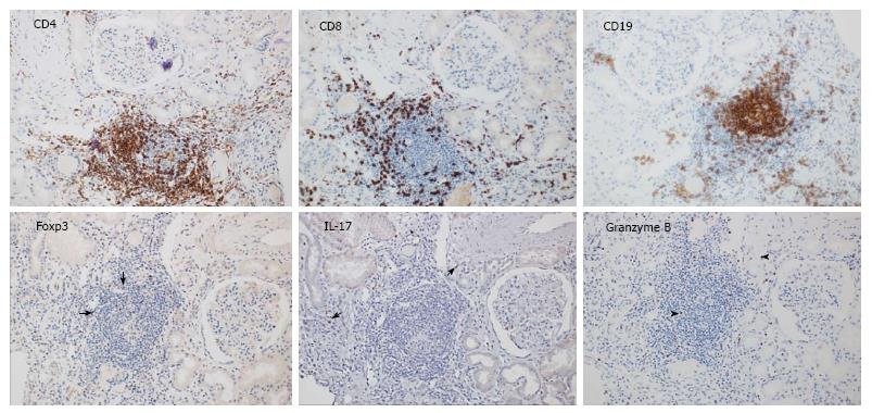
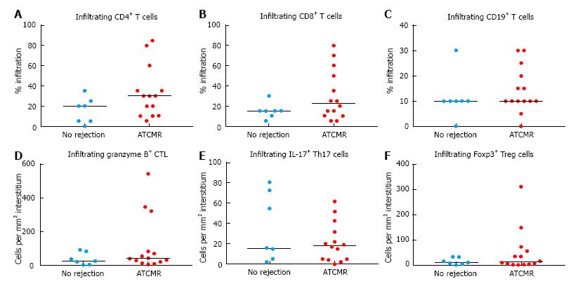
Figure 1 shows a representative panel of the immunohostochemical analysis of T cell subsets in a patient with ATCMR-KTx. The percentage of CD4+ and CD8+ cell infiltration was higher in patients with ATCMR-KTx (Figure 2A and B), and there was no significant difference in B cell infiltration (Figure 2C) in comparison to patients with no rejection. The infiltration of granzyme B+ cells (surrogates for CTL), Foxp3+ cells (surrogates for Treg cells) and IL-17+ cells (surrogates for Th17 cells), quantified as cell densities (number of cells per mm2 of tubulo-interstitial biopsy area), were not statistically different between the two patient groups (Figure 2D-F). Nonetheless, a few ATCMR-KTx patients had higher infiltration by granzyme B+ and Foxp3+ cells and are referred subsequently in the text as ‘high infiltration outliers’.
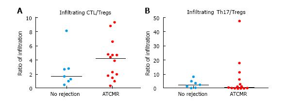
As an arbitrary measurement of immune balance within the kidney transplant, the granzyme B+ cell to Foxp3+ cell density ratio was found to be higher in patients with ATCMR-KTx than for patients in which rejection was not observed (Figure 3A). However, the ratio of infiltrating IL-17-producing cells over Foxp3+ cells was not much different in patients with ATCMR-KTx than in patients not experiencing rejection (Figure 3B). Given our small sample size, these comparisons did not achieve statistical significance. However, once more there were a few “high infiltration outliers” for the ratio of infiltrating Th17 cells over Foxp3+ Treg cells.
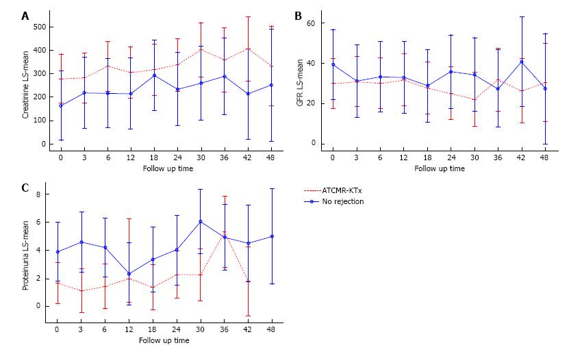
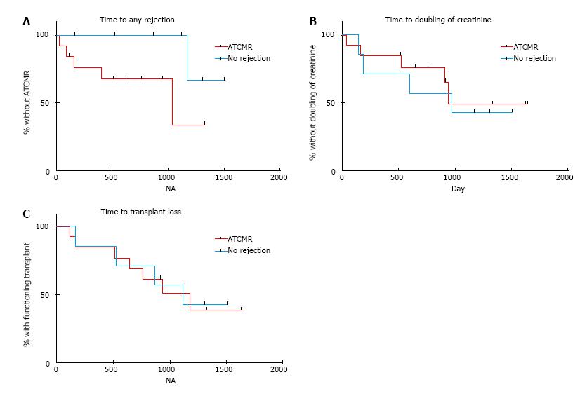
The numbers of infiltrating Th17 cells in the ATCMR-KTx patients were significantly positively correlated with serum creatinine levels and proteinuria, and negatively correlated with eGFR at different time points during follow up. The numbers of infiltrating Th17 cells and the ratio of Th17 cells over Foxp3+ Treg cells in the non-rejection patients were significantly positively correlated with serum creatinine levels and negatively correlated with eGFR at different time points during follow up. Correlation estimates and P values of the statistically significant associations are shown in Table 4. The numbers of infiltrating CTL and infiltrating Foxp3+ Treg cells were not significantly associated with any of the clinical outcomes tested including changes in serum creatinine, eGFR or proteinuria. However, a significant negative correlation of the ratio of infiltrating CTL over Foxp3+ Tregs with creatinine at 3 mo was observed in ATCMR-KTx patients. Figure 4 shows the dynamic changes in serum creatinine, eGFR and proteinuria throughout the follow up period. The ATCMR-KTx group had overall worse kidney transplant function during follow up than the non-rejection group, while the non-rejection group had overall higher levels of proteinuria. There was no more rapid deterioration in the ATCMR-KTx patients in comparison to the non-rejection patients, as indicated by the absence of statistically significant differences between respective mean values for changes in serum creatinine, eGFR and proteinuria. The time-to-event plots for any rejection post-biopsy (borderline, ATCMR-KTx or antibody-mediated rejection), time to doubling of creatinine post-biopsy, and time to confirmed or suspected immune-mediated transplant loss are found in Figure 5. Table 5 contains the respective median times to event. The comparisons of the time-to-event curves by log rank test were not statistically significant. The effect of the cell densities of the infiltrating immune cells and their ratios, as well as the effect of clinical parameters suspected to influence kidney transplant outcomes (i.e., the potential confounders for kidney transplant outcomes taken from Table 1) were tested using cox regression model. Their respective hazard ratios and 95%CI are shown in Table 6. In the univariate analysis, younger age was associated significantly with shorter time to any rejection. In addition, the number of infiltrating Th17 cells and the degree of proteinuria at biopsy were significantly associated with shorter time to doubling of creatinine. The number of infiltrating Th17 cells, serum creatinine at biopsy and the occurrence of delayed graft function were significantly associated with shorter time to transplant loss. Multivariate analysis was not performed in consequence of the small sample size.
| Group | Immune parameter | vs | Outcome | R | P value |
| No rejection | Infiltrating Th17 cells | Creatinine t3 | 0.9429 | 0.0167 | |
| No rejection | Infiltrating Th17 cells | GFR t0 | -0.8571 | 0.0238 | |
| No rejection | Infiltrating Th17/Tregs | GFR t0 | -0.7857 | 0.048 | |
| No rejection | Infiltrating Th17 cells | GFR t3 | -0.9429 | 0.0167 | |
| No rejection | Infiltrating Th17/Tregs | GFR t3 | -0.9429 | 0.0167 | |
| No rejection | Infiltrating Th17 cells | GFR t6 | -0.8929 | 0.0123 | |
| ATCMR-KTx | Infiltrating CTL/Tregs | Creatinine t3 | -0.6694 | 0.0145 | |
| ATCMR-KTx | Infiltrating Th17 cells | Creatinine t24 | 0.6485 | 0.049 | |
| ATCMR-KTx | Infiltrating Th17 cells | Creatinine t30 | 0.7619 | 0.0368 | |
| ATCMR-KTx | Infiltrating Th17 cells | GFR t30 | -0.8333 | 0.0154 | |
| ATCMR-KTx | Infiltrating Th17 cells | Proteinuria t12 | 0.8095 | 0.0218 |
| Outcomes | Group | Median time-to-event | P values |
| Any rejection | ATCMR | 1037 | 0.0941 |
| No rejection | Undefined1 | ||
| Doubling of creatinine | ATCMR | 941 | 0.7452 |
| No rejection | 974 | ||
| Transplant loss | ATCMR | 1176 | 0.956 |
| No rejection | 1118 |
| Outcomes | Risk factor | HR | 95%CI | P value |
| Time to any rejection | Age | 0.898 | 0.821, 0.983 | 0.0193 |
| Time to doubling of creatinine | Infiltrating Th17 cells | 1.031 | 1.002, 1.061 | 0.0359 |
| Time to doubling of creatinine | Proteinuria | 1.382 | 1.087, 1.757 | 0.0083 |
| Time to transplant loss | Infiltrating Th17 cells | 1.026 | 1.000, 1.052 | 0.0472 |
| Time to transplant loss | Serum creatinine | 1.009 | 1.003, 1.016 | 0.0036 |
| Time to transplant loss | Delayed graft function | 5.456 | 1.238, 24.036 | 0.0160 |
Finally, for ATCMR-KTx patients, Kaplan-Meier time-to-event curves for kidney transplant outcomes corresponding to “high infiltration outlier” patients were compared to outcomes for “non-outlier” patients relative to: (1) number of infiltrating CTL; (2) number of infiltrating Foxp3+ Treg cells; and (3) ratio of Th17 cell to Foxp3+ Treg cell. Owing to the small sample sizes, median time-to-event was not obtainable for any outcome, and differences between “outlier” and “non-outlier” survival curves were non-significant for all three outcome variables (data not shown).
In this study, our main aim was to determine whether the T cell subset composition in ATCMR-KTx differed qualitatively or quantitatively from that in the absence of rejection. Our main focus was on the numbers and respective ratios of CTL, Th17 cells and Foxp3+ Treg cells, thought to be the most relevant subsets implicated in ATCMR-KTx, according the previously presented literature. ATCMR-KTx appeared to be characterised by a numerical dominance of CTL over Foxp3+ Treg cells in comparison to the absence of acute rejection, suggesting that the immune balance in ATCMR-KTx appears to be tilted to the pro-rejection forces; which might be overwhelming the regulatory forces. This finding is congruent with the literature reports, where the presence of CTL infiltrating the kidney transplant undergoing ATCMR is a characteristic to differentiate ATCMR-KTx from the absence of rejection[10,12,13]; and with the published observation that a lower infiltration by Foxp3+ Treg cells in the kidney transplant undergoing ATCMR was associated with poorer transplant outcomes[26], or with poorer responsiveness to anti-rejection therapy[20].
Our analysis of kidney transplant outcomes revealed that the number of infiltrating Th17 cells was significantly associated with faster time to doubling of creatinine and transplant loss; and the ratio of infiltrating Th17 cells over Foxp3+ Treg cells was significantly associated with a decline in eGFR. These findings parallel and further support the published observations where the magnitude of Th17 cell infiltration over Treg cell infiltration correlated with kidney transplant dysfunction, the degree of interstitial inflammation and tubular atrophy, the refractoriness to treatment and the recurrence of ATCMR in the kidney transplant[21,22]. However, the associations observed in our study were not very strong. The observation that the numbers of infiltrating Th17 cells and the ratio of Th17 cells over Foxp3+ Treg cells associated negatively with kidney transplant outcomes in the non-rejection patients was unexpected, but interesting. Alloimmune responses in transplant patients are detectable even in patients with apparent stable kidney function. Different sorts of immune cells are as a consequence “waiting for a chance” to flip over the silencing effects of maintenance immunosuppression and the deployed immunoregulatory mechanisms if “given the chance” (i.e., reduction of immunosuppression, sensitizing events or the occurrence of concomitant infections or inflammatory disorders). Hence, it is possible that may transplant patients have certain degree of Th17 cell activation and infiltration. Thus, patients with higher degree of Th17 infiltration, irrespective of reaching the current thresholds for ATCMR-KTx or not, could be bound for worse outcomes due to the possibility that Th17 cells could be mediating smoldering inflammation or slow-motion chronic rejection or have the potential to mediate transformation into a rejection phenotype if the alloimmune milieu changes to a pro-inflammatory one. The use of more sophisticated technologies like the molecular microscope and a better classification of chronic T cell mediated rejection and i-IFTA (for inflamed areas of interstitial fibrosis and tubular atrophy) could help us in the future to assign a more accurate clinical significance to this interesting observation.
In contrast to published literature, in which a greater degree of infiltration by CTL in patients with ATCMR-KTx was associated with poorer allograft survival[18], and the magnitude of granzyme B expression was associated with the severity of the rejection process[10]; we found no association of CTL infiltration or the ratio of infiltrating CTL over Treg cells with kidney transplant outcomes. However, we believe that statistical significance was not reached due to our small pilot sample size.
One of the merits of our study is the use of immunohistochemistry for our immunodetection as it is a highly available and inexpensive technology, easy to correlate to conventional light microscopy findings. Furthermore, in comparison to most available reports, our study provides a more comprehensive tissue staining, including the three markers that showed the best potential in the published literature: Granzyme B, IL-17 and Foxp3. Thus, our study hints that a more detailed immunohistochemical analysis of the cell infiltrate in kidney transplant biopsies can reflect more accurately the immune balance between the pro-rejection and anti-rejection forces and opens avenues for larger more powered and comprehensive confirmatory studies to address whether a detailed immunophenotyping of ATCMR-KTx can indeed improve the accuracy of the Banff classification; which is undergoing continuous improvement. It is important to comment that more sophisticated technologies like microarray technology have been used for the detection of CTL-associated transcripts and were reported to be more accurate than the detection of individual genes like perforin or granzyme B to cluster together patients with ATCMR-KTx[31]. However, this latter technology is not widely available and not as practical as immunohistochemistry; but indeed, microarray and high-throughput technologies such as the “omics” play a crucial role in biomarker discovery and identification of disease classifiers.
In addressing sample size, based upon our pilot study results, assuming a 1:2 sample size ratio of non-rejection:ATCMR-KTx patients, a common standard deviation (σ) and coefficient of variation (CV = σ/μNoReject) 1.0 to 1.7, respective optimistic and pessimistic sample sizes to give 80% power to detect a two-fold ratio of CTL (CTL: Non-rejection/ATCMR-KTx ≥ 2) to Foxp3+ Treg cells were calculated to be 18/36 (CV = 1.0) and 41/82 (CV = 1.7).
Participating patients were very heterogeneous in their clinical characteristics, which likely confounded our observations (Tables 1, 2 and 3). For instance, we observed that the time to transplant loss from biopsy (not from transplant surgery) was similar in both patient groups. However, most kidney transplant biopsies in the non-rejection group were performed late post-transplantation, closer to their maximum transplant survival. In addition, the non-rejection group had higher proteinuria during the follow up period, which could be related also to their vintage in transplantation and likely higher degree of glomerulosclerosis, or perhaps proteinuria was an important factor in the decision to perform biopsy for those patients. Kidney transplant biopsies were indicated when transplant dysfunction ensued and recommended by treating nephrologists according to their own criteria and specific thresholds. The incorporation of selected immune parameters in a larger study including patients from the time of transplant surgery, subjected to more protocolised immunosuppressive regimens, or their incorporation in a clinical trial are anticipated to circumvent many of the biases in our study.
Finally, it would have been interesting to extend our protocol to assess the immune infiltrate inside the kidney transplant in protocol biopsies with subclinical ATCMR and without evidence of rejection. This could have helped us to address whether our observed immune changes mirror the events occurring in subclinical ATCMR-KTx, and to use negative protocol biopsies as better controls for a stable kidney transplant function. However, protocol biopsies are not performed in our institution.
The immune balance in ATCMR-KTx appears to be tilted numerically towards the pro-rejection forces, which seem to overwhelm counter-regulatory mechanisms. Similarly, the degree of infiltration of the kidney transplant by effector T cells could be associated with kidney transplant outcome prognosis. Although our findings are not conclusive, mainly due to our small sample size, they further elucidate the immunopathogenesis of ATCMR-KTx and open new avenues for a more detailed dissection of the complex immune mechanisms implicated in kidney transplant rejection. Upon further validation, ideally tested in randomised controlled trials, it is possible that these and other new signatures could be incorporated into the current diagnostic and therapeutic algorithms in order to deliver more personalised and precise management in kidney transplantation.
We would like to acknowledge the Department of Clinical Research of the Singapore General Hospital for allowing to use their computers for data collection. One thousand thank-you’s to Madam Rachel Liew, our library technician, for helping us obtain some of the least accessible articles; and to the anonymous reviewers of the journal for their useful comments. We are also much obliged to the National Kidney Foundation Singapore; the Medicine Academic Clinical Program (a SingHealth-Duke/National University of Singapore Joint Partnership); and the Khoo Scholar Programme (Duke/National University of Singapore) for generously funding different aspects of our research on T cell subset analysis in kidney transplantation.
In the clinical setting, acute T cell-mediated rejection in the kidney transplant (ATCMR-KTx) is only confirmed through a kidney transplant biopsy, which is scored according to the Banff classification. The Banff classification is largely based on the estimation of mononuclear cell infiltration instead of the identification and quantification of the actual T cell subsets recruited to mediate rejection.
The identification of the actual T cell subsets involved in ATCMR-KTx likely reflects more accurately the immune balance between effector and regulatory T cells, which has been implicated as an important factor determining the risk for ATCMR-KTx.
The detection of specific T cell subsets inside the kidney transplant suffering ATCMR adds new light to elucidate its immunopathogenesis, and opens new avenues for the development of novel biomarkers focusing on cytotoxic, Th17 cell-mediated and regulatory T cell responses.
A more detailed analysis of the inflammatory infiltrate of ATCMR-KTx, in particular of cytotoxic T lymphocytes and Th17 cells, is likely to enhance the diagnostic accuracy of the Banff classification.
CD178: CD equivalent for Fas ligand, a membrane molecule able to trigger apoptosis upon ligation of CD95 in target allogeneic cells; Cytotoxic T lymphocytes: A subset of effector T cells able to cause direct cytotoxicity of transplanted parenchymal cells; Foxp3: Transcription factor crucial for the development and function of regulatory T cells; Granzyme B: Enzyme released by cytotoxic T lymphocytes able to trigger apoptosis in target transplanted cells; Regulatory T cells: A subset of T cells regarded as the master moderators of immune responses, thought to be able to regulate alloimmune responses and potentially to aid in the achievement of transplantation tolerance; Th17 cells: A subset of effector T cells implicated in the defence against exogenous microorganisms and implicated in the pathogenesis of several autoimmune disorders and effector alloresponses, whose characteristic cytokine product is IL-17.
This is a good article.
Manuscript source: Invited manuscript
Specialty type: Transplantation
Country of origin: Singapore
Peer-review report classification
Grade A (Excellent): 0
Grade B (Very good): B
Grade C (Good): C
Grade D (Fair): 0
Grade E (Poor): 0
P- Reviewer: Salvadori M, Taheri S S- Editor: Ji FF L- Editor: A E- Editor: Lu YJ
| 1. | Opelz G, Döhler B. Influence of time of rejection on long-term graft survival in renal transplantation. Transplantation. 2008;85:661-666. [PubMed] [DOI] [Cited in This Article: ] [Cited by in Crossref: 89] [Cited by in F6Publishing: 100] [Article Influence: 6.3] [Reference Citation Analysis (0)] |
| 2. | Schnuelle P, Lorenz D, Trede M, Van Der Woude FJ. Impact of renal cadaveric transplantation on survival in end-stage renal failure: evidence for reduced mortality risk compared with hemodialysis during long-term follow-up. J Am Soc Nephrol. 1998;9:2135-2141. [PubMed] [Cited in This Article: ] |
| 3. | Loupy A, Haas M, Solez K, Racusen L, Glotz D, Seron D, Nankivell BJ, Colvin RB, Afrouzian M, Akalin E. The Banff 2015 Kidney Meeting Report: Current Challenges in Rejection Classification and Prospects for Adopting Molecular Pathology. Am J Transplant. 2017;17:28-41. [PubMed] [DOI] [Cited in This Article: ] [Cited by in Crossref: 447] [Cited by in F6Publishing: 482] [Article Influence: 68.9] [Reference Citation Analysis (0)] |
| 4. | Benghiat FS, Graca L, Braun MY, Detienne S, Moore F, Buonocore S, Flamand V, Waldmann H, Goldman M, Le Moine A. Critical influence of natural regulatory CD25+ T cells on the fate of allografts in the absence of immunosuppression. Transplantation. 2005;79:648-654. [PubMed] [DOI] [Cited in This Article: ] [Cited by in Crossref: 69] [Cited by in F6Publishing: 70] [Article Influence: 3.7] [Reference Citation Analysis (0)] |
| 5. | Chai JG, Xue SA, Coe D, Addey C, Bartok I, Scott D, Simpson E, Stauss HJ, Hori S, Sakaguchi S. Regulatory T cells, derived from naïve CD4+CD25- T cells by in vitro Foxp3 gene transfer, can induce transplantation tolerance. Transplantation. 2005;79:1310-1316. [PubMed] [DOI] [Cited in This Article: ] [Cited by in Crossref: 108] [Cited by in F6Publishing: 112] [Article Influence: 5.9] [Reference Citation Analysis (0)] |
| 6. | Joffre O, Santolaria T, Calise D, Al Saati T, Hudrisier D, Romagnoli P, van Meerwijk JP. Prevention of acute and chronic allograft rejection with CD4+CD25+Foxp3+ regulatory T lymphocytes. Nat Med. 2008;14:88-92. [PubMed] [DOI] [Cited in This Article: ] [Cited by in Crossref: 402] [Cited by in F6Publishing: 427] [Article Influence: 25.1] [Reference Citation Analysis (0)] |
| 7. | Zheng XX, Sanchez-Fueyo A, Domenig C, Strom TB. The balance of deletion and regulation in allograft tolerance. Immunol Rev. 2003;196:75-84. [PubMed] [DOI] [Cited in This Article: ] [Cited by in Crossref: 85] [Cited by in F6Publishing: 88] [Article Influence: 4.4] [Reference Citation Analysis (0)] |
| 8. | Graca L, Cobbold SP, Waldmann H. Identification of regulatory T cells in tolerated allografts. J Exp Med. 2002;195:1641-1646. [PubMed] [DOI] [Cited in This Article: ] [Cited by in Crossref: 458] [Cited by in F6Publishing: 451] [Article Influence: 20.5] [Reference Citation Analysis (0)] |
| 9. | Hu M, Wang C, Zhang GY, Saito M, Wang YM, Fernandez MA, Wang Y, Wu H, Hawthorne WJ, Jones C. Infiltrating Foxp3(+) regulatory T cells from spontaneously tolerant kidney allografts demonstrate donor-specific tolerance. Am J Transplant. 2013;13:2819-2830. [PubMed] [DOI] [Cited in This Article: ] [Cited by in Crossref: 48] [Cited by in F6Publishing: 49] [Article Influence: 4.5] [Reference Citation Analysis (0)] |
| 10. | Wagrowska-Danilewicz M, Danilewicz M. Immunoexpression of perforin and granzyme B on infiltrating lymphocytes in human renal acute allograft rejection. Nefrologia. 2003;23:538-544. [PubMed] [Cited in This Article: ] |
| 11. | Rascio F, Divella C, Grandaliano G. CTL and transplantation: tissue in vivo characterization. Methods Mol Biol. 2014;1186:283-294. [PubMed] [DOI] [Cited in This Article: ] [Cited by in Crossref: 1] [Cited by in F6Publishing: 2] [Article Influence: 0.2] [Reference Citation Analysis (0)] |
| 12. | Ashton-Chess J, Dugast E, Colvin RB, Giral M, Foucher Y, Moreau A, Renaudin K, Braud C, Devys A, Brouard S. Regulatory, effector, and cytotoxic T cell profiles in long-term kidney transplant patients. J Am Soc Nephrol. 2009;20:1113-1122. [PubMed] [DOI] [Cited in This Article: ] [Cited by in Crossref: 54] [Cited by in F6Publishing: 56] [Article Influence: 3.7] [Reference Citation Analysis (0)] |
| 13. | Desvaux D, Schwarzinger M, Pastural M, Baron C, Abtahi M, Berrehar F, Lim A, Lang P, le Gouvello S. Molecular diagnosis of renal-allograft rejection: correlation with histopathologic evaluation and antirejection-therapy resistance. Transplantation. 2004;78:647-653. [PubMed] [DOI] [Cited in This Article: ] [Cited by in Crossref: 40] [Cited by in F6Publishing: 43] [Article Influence: 2.2] [Reference Citation Analysis (0)] |
| 14. | Heng B, Li Y, Shi L, Du X, Lai C, Cheng L, Su Z. A Meta-analysis of the Significance of Granzyme B and Perforin in Noninvasive Diagnosis of Acute Rejection After Kidney Transplantation. Transplantation. 2015;99:1477-1486. [PubMed] [DOI] [Cited in This Article: ] [Cited by in Crossref: 16] [Cited by in F6Publishing: 18] [Article Influence: 2.3] [Reference Citation Analysis (0)] |
| 15. | Sabek O, Dorak MT, Kotb M, Gaber AO, Gaber L. Quantitative detection of T-cell activation markers by real-time PCR in renal transplant rejection and correlation with histopathologic evaluation. Transplantation. 2002;74:701-707. [PubMed] [DOI] [Cited in This Article: ] [Cited by in Crossref: 78] [Cited by in F6Publishing: 80] [Article Influence: 3.6] [Reference Citation Analysis (0)] |
| 16. | Graziotto R, Del Prete D, Rigotti P, Anglani F, Baldan N, Furian L, Valente M, Antonello A, Marchini F, D’Angelo A. Perforin, Granzyme B, and fas ligand for molecular diagnosis of acute renal-allograft rejection: analyses on serial biopsies suggest methodological issues. Transplantation. 2006;81:1125-1132. [PubMed] [DOI] [Cited in This Article: ] [Cited by in Crossref: 34] [Cited by in F6Publishing: 37] [Article Influence: 2.1] [Reference Citation Analysis (0)] |
| 17. | Sarwal MM, Jani A, Chang S, Huie P, Wang Z, Salvatierra O, Clayberger C, Sibley R, Krensky AM, Pavlakis M. Granulysin expression is a marker for acute rejection and steroid resistance in human renal transplantation. Hum Immunol. 2001;62:21-31. [PubMed] [DOI] [Cited in This Article: ] [Cited by in Crossref: 66] [Cited by in F6Publishing: 71] [Article Influence: 3.1] [Reference Citation Analysis (0)] |
| 18. | Mengel M, Mueller I, Behrend M, von Wasielewski R, Radermacher J, Schwarz A, Haller H, Kreipe H. Prognostic value of cytotoxic T-lymphocytes and CD40 in biopsies with early renal allograft rejection. Transpl Int. 2004;17:293-300. [PubMed] [DOI] [Cited in This Article: ] [Cited by in F6Publishing: 3] [Reference Citation Analysis (0)] |
| 19. | Nickel P, Lacha J, Ode-Hakim S, Sawitzki B, Babel N, Frei U, Volk HD, Reinke P. Cytotoxic effector molecule gene expression in acute renal allograft rejection: correlation with clinical outcome; histopathology and function of the allograft. Transplantation. 2001;72:1158-1160. [PubMed] [DOI] [Cited in This Article: ] [Cited by in Crossref: 33] [Cited by in F6Publishing: 35] [Article Influence: 1.5] [Reference Citation Analysis (0)] |
| 20. | Matignon M, Aissat A, Canoui-Poitrine F, Grondin C, Pilon C, Desvaux D, Saadoun D, Barathon Q, Garrido M, Audard V. Th-17 Alloimmune Responses in Renal Allograft Biopsies From Recipients of Kidney Transplants Using Extended Criteria Donors During Acute T Cell-Mediated Rejection. Am J Transplant. 2015;15:2718-2725. [PubMed] [DOI] [Cited in This Article: ] [Cited by in Crossref: 17] [Cited by in F6Publishing: 19] [Article Influence: 2.1] [Reference Citation Analysis (0)] |
| 21. | Chung BH, Oh HJ, Piao SG, Sun IO, Kang SH, Choi SR, Park HS, Choi BS, Choi YJ, Park CW. Higher infiltration by Th17 cells compared with regulatory T cells is associated with severe acute T-cell-mediated graft rejection. Exp Mol Med. 2011;43:630-637. [PubMed] [DOI] [Cited in This Article: ] [Cited by in Crossref: 19] [Cited by in F6Publishing: 22] [Article Influence: 1.8] [Reference Citation Analysis (0)] |
| 22. | Chung BH, Oh HJ, Piao SG, Hwang HS, Sun IO, Choi SR, Park HS, Choi BS, Choi YJ, Park CW. Clinical significance of the ratio between FOXP3 positive regulatory T cell and interleukin-17 secreting cell in renal allograft biopsies with acute T-cell-mediated rejection. Immunology. 2012;136:344-351. [PubMed] [DOI] [Cited in This Article: ] [Cited by in Crossref: 30] [Cited by in F6Publishing: 32] [Article Influence: 2.7] [Reference Citation Analysis (0)] |
| 23. | Wood KJ, Sakaguchi S. Regulatory T cells in transplantation tolerance. Nat Rev Immunol. 2003;3:199-210. [PubMed] [DOI] [Cited in This Article: ] [Cited by in Crossref: 1059] [Cited by in F6Publishing: 1012] [Article Influence: 48.2] [Reference Citation Analysis (0)] |
| 24. | Louis S, Braudeau C, Giral M, Dupont A, Moizant F, Robillard N, Moreau A, Soulillou JP, Brouard S. Contrasting CD25hiCD4+T cells/FOXP3 patterns in chronic rejection and operational drug-free tolerance. Transplantation. 2006;81:398-407. [PubMed] [DOI] [Cited in This Article: ] [Cited by in Crossref: 221] [Cited by in F6Publishing: 231] [Article Influence: 12.8] [Reference Citation Analysis (0)] |
| 25. | Salcido-Ochoa F, Yusof N, Hue SS, Haase D, Kee T, Rotzschke O. Are we ready for the use of foxp3(+) regulatory T cells for immunodiagnosis and immunotherapy in kidney transplantation? J Transplant. 2012;2012:397952. [PubMed] [DOI] [Cited in This Article: ] [Cited by in Crossref: 3] [Cited by in F6Publishing: 5] [Article Influence: 0.4] [Reference Citation Analysis (0)] |
| 26. | Martin L, Funes de la Vega M, Bocrie O, Harzallah A, Justrabo E, Rifle G, Mousson C. Detection of Foxp3+ cells on biopsies of kidney transplants with early acute rejection. Transplant Proc. 2007;39:2586-2588. [PubMed] [DOI] [Cited in This Article: ] [Cited by in Crossref: 11] [Cited by in F6Publishing: 10] [Article Influence: 0.6] [Reference Citation Analysis (0)] |
| 27. | Bestard O, Cruzado JM, Mestre M, Caldés A, Bas J, Carrera M, Torras J, Rama I, Moreso F, Serón D. Achieving donor-specific hyporesponsiveness is associated with FOXP3+ regulatory T cell recruitment in human renal allograft infiltrates. J Immunol. 2007;179:4901-4909. [PubMed] [DOI] [Cited in This Article: ] [Cited by in Crossref: 122] [Cited by in F6Publishing: 132] [Article Influence: 7.8] [Reference Citation Analysis (0)] |
| 28. | Bestard O, Cuñetti L, Cruzado JM, Lucia M, Valdez R, Olek S, Melilli E, Torras J, Mast R, Gomà M. Intragraft regulatory T cells in protocol biopsies retain foxp3 demethylation and are protective biomarkers for kidney graft outcome. Am J Transplant. 2011;11:2162-2172. [PubMed] [DOI] [Cited in This Article: ] [Cited by in Crossref: 67] [Cited by in F6Publishing: 65] [Article Influence: 5.0] [Reference Citation Analysis (0)] |
| 29. | Kollins D, Stoelcker B, Hoffmann U, Bergler T, Reinhold S, Banas MC, Rümmele P, Farkas S, Krämer BK, Banas B. FOXP3+ regulatory T-cells in renal allografts: correlation with long-term graft function and acute rejection. Clin Nephrol. 2011;75:91-100. [PubMed] [Cited in This Article: ] |
| 30. | Fuggle SV, Belger MA, Johnson RJ, Ray TC, Morris PJ. A new national allocation scheme for adult kidneys in the United Kingdom. United Kingdom Transplant Support Service Authority (UKTSSA) Users’ Kidney Advisory Group and its Task Forces. Clin Transpl. 1998;107-113. [PubMed] [Cited in This Article: ] |
| 31. | Hidalgo LG, Einecke G, Allanach K, Mengel M, Sis B, Mueller TF, Halloran PF. The transcriptome of human cytotoxic T cells: measuring the burden of CTL-associated transcripts in human kidney transplants. Am J Transplant. 2008;8:637-646. [PubMed] [DOI] [Cited in This Article: ] [Cited by in Crossref: 56] [Cited by in F6Publishing: 57] [Article Influence: 3.6] [Reference Citation Analysis (0)] |


