Published online Apr 28, 2013. doi: 10.4329/wjr.v5.i4.184
Revised: December 30, 2012
Accepted: January 31, 2013
Published online: April 28, 2013
We present a case of the right aortic arch with kommerell diverticulum (KD) and aberrant left subclavian artery in a symptomatic 50-year-old patient with a calcification in the presumed attachment site of the ligamentum arteriosum (LA) to the KD. In another 30-year-old male patient, the entire course of a calcified LA was demonstrated using multidetector row computed tomography.
- Citation: Kanza RE, Berube M, Michaud P. MDCT of right aortic arch with aberrant left subclavian artery associated with kommerell diverticulum and calcified ligamentum arteriosum. World J Radiol 2013; 5(4): 184-186
- URL: https://www.wjgnet.com/1949-8470/full/v5/i4/184.htm
- DOI: https://dx.doi.org/10.4329/wjr.v5.i4.184
Right aortic arch with aberrant origin of left subclavian artery is rare congenital variation of the aortic arch and its branches with a reported prevalence of 0.05%-1% in the literature[1]. This anomaly is frequently an incidental findings during autopsies series or angiographic studies because it is usually asymptomatic, but rarely may be symptomatic especially when there is a partial or complete obstruction of the oesophagus and/or trachea.
With the recent development of newer non-invasive imaging systems, the use computed tomography and magnetic resonance imaging (MRI) in daily practice has been increase to diagnose and characterize anomalies of aortic arch and its branches including vascular ring. Despite this, the last part or the complete vascular ring which is made by left-side ligamentum arteriosum is often not clearly visualized pre-operatively.
We present multidetector row computed tomography (MDCT)-angiographic findings of a case of right aortic arch (RAA) with aberrant left subclavian artery (ALSA) associated with Kommerell diverticulum and calcified ligamentum arteriosum causing dysphagia lusoria. In another patient, we emphasize the role that MDCT may play to demonstrate calcified ligamentous arteriosum (LA).
A 50-year-old man was referred to our department for investigation of dysphagia.
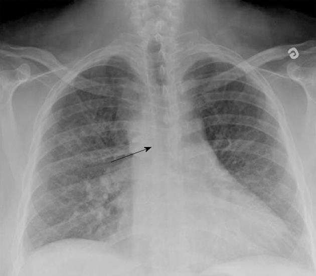
The patient had a long history of arterial hypertension, type II diabetes mellitus, and sleep apnea syndrome and was newly diagnosed with ischemic cardiac disease. A chest X-ray showed a right aortic knob and arch with mild tracheal deviation to the left (Figure 1).
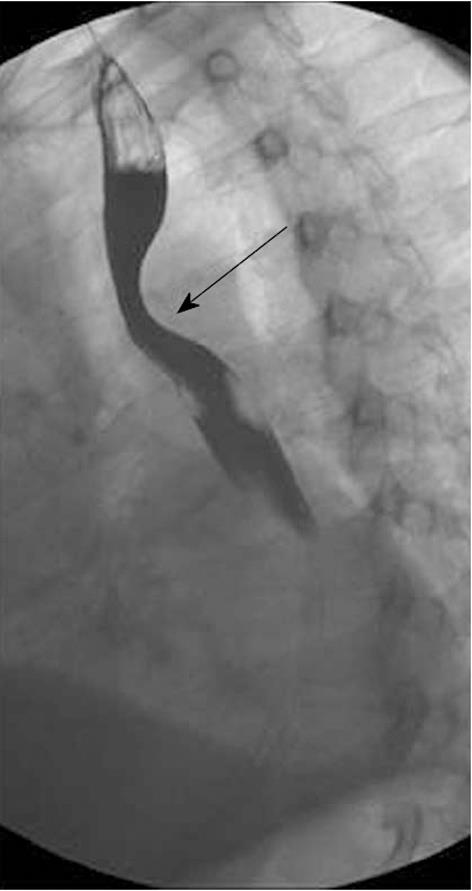
A barium esophagogram showed posterior compression of the esophagus (Figure 2).
MDCT-angiography was performed using a 16-slice MDCT scanner (LightSpeed 16, GE Healthcare, WI, Milwaukee, United States). CT parameters included 2.5-mm slice thickness, gantry rotation time 0.5 s, 120 Kvp and 300 mA. The scanning delay was determined with a bolus tracking technique. The true CT study was obtained after administration of 120 cc of contrast medium at the rate of 4 cc/s. Multiplanar, maximum intensity projection and three dimensional volume-rendered reformations were obtained using a separate workstation (Voxar 3D in impax 6.3; Agfa).
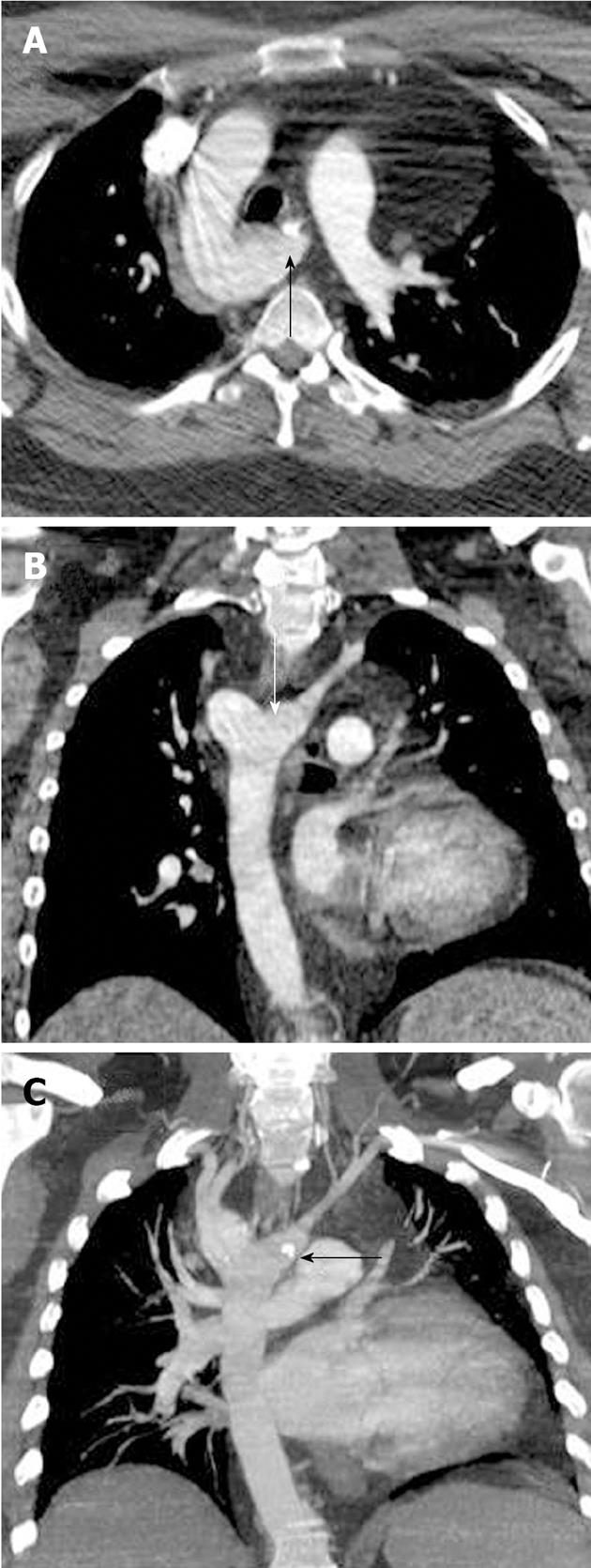
MDCT-angiography demonstrated RAA with a kommerell diverticulum (KD) and an ALSA (Figure 3A and B). Additionally, calcification was found in the presumed site of the superior attachment of LA to the KD (Figure 3C).
Because of multiple comorbidities and relatively non-disabling dysphagia, surgery was postponed and priority was given to thoroughly investigating the patient’s cardiac disease and controlling all the comorbities before safe surgery could be planned.
A 30-year-old referred to our department for chest CT following detection of 6 mm lung nodule in the right lung base during abdominal CT performed for abdominal pain investigation. The chest CT shows calcification of the entire course of the ligamentum arteriosum as an incidental findings (Figure 4)
Right aortic arch with aberrant origin of left subclavian artery is one of the commonest mediastinal vascular ring. This anomaly is often asymptomatic and most of patient will remain symptom-free in their life time. Rarely patients may present symptoms in early life time or become symptomatic in young adulthood or later due the compression of the oesophagus and/or trachea leading to dysphagia and/or dyspnoea. Surgery may be required if symptoms are moderate or severe. We present a new case of RAA with ALSA and KD causing adult onset of dysphagia lusoria. The diagnosis was suggested by the chest X-ray and barium esophagram but confirmed by MDCT-angiophaphy with better characterization of the vascular ring.
This case illustrates the role of MDCT-angiography in the diagnosis of RAA with KD and ALSA. This role is quite similar with those of MRI. Although CT and MRI are very useful for the diagnosis of a vascular ring in routine clinical practice, the last part of the ring in a complete vascular ring, which is made by the LA, is often not clearly seen preoperatively, to guide surgical division. Several recent studies have shown that with modern imaging techniques, especially CT, the rate at which LA calcification is being detected has increased, varying from 11.2% to 48%[2,3]. Calcification usually occurs at the aortic end of the LA - or at the KD near the take-off of the ALSA; it may even show patterns, including curvilinear, clumped, punctuate or linear[3] (Figure 4).
In their recent article, Paparo et al[4] show that MRI could be a powerful imaging tool for the diagnosis of vascular rings and may be useful for demonstrating the course of the LA. However, we believe in case of LA calcification, MDCT may be superior to MRI. Further, MDCT has the advantage of being fast and widely available, while MRI may not be suitable for patients with claustrophobia, cardiac pacemakers, significant dyspnea, or the need for sedation. The main drawback of CT remains the radiation dose and the use of a contrast medium. In routine clinical practice, the choice of modality may depend mainly on the patient’s age, type of surgery (open vs endoscopic), and expertise at the institution where the surgery is to be conducted.
P- Reviewer Yazdi HR S- Editor Cheng JX L- Editor A E- Editor Xiong L
| 1. | Cinà CS, Althani H, Pasenau J, Abouzahr L. Kommerell’s diverticulum and right-sided aortic arch: a cohort study and review of the literature. J Vasc Surg. 2004;39:131-139. [PubMed] [DOI] [Cited in This Article: ] [Cited by in Crossref: 254] [Cited by in F6Publishing: 246] [Article Influence: 12.3] [Reference Citation Analysis (0)] |
| 2. | Hong GS, Goo HW, Song JW. Prevalence of ligamentum arteriosum calcification on multi-section spiral CT and digital radiography. Int J Cardiovasc Imaging. 2012;28 Suppl 1:61-67. [PubMed] [Cited in This Article: ] |
| 3. | Wimpfheimer O, Haramati LB, Haramati N. Calcification of the ligamentum arteriosum in adults: CT features. J Comput Assist Tomogr. 1996;20:34-37. [PubMed] [DOI] [Cited in This Article: ] [Cited by in Crossref: 13] [Cited by in F6Publishing: 15] [Article Influence: 0.5] [Reference Citation Analysis (0)] |
| 4. | Paparo F, Bacigalupo L, Melani E, Rollandi GA, Caro GD. Cardiac-MRI demonstration of the ligamentum arteriosum in a case of right aortic arch with aberrant left subclavian artery. World J Radiol. 2012;4:231-235. [PubMed] [DOI] [Cited in This Article: ] [Cited by in CrossRef: 3] [Cited by in F6Publishing: 4] [Article Influence: 0.3] [Reference Citation Analysis (0)] |


