Published online Jun 15, 1999. doi: 10.3748/wjg.v5.i3.270
Revised: February 22, 1999
Accepted: March 13, 1999
Published online: June 15, 1999
- Citation: Huang RT, Li XY, Xia XB, Yuan XT, Liu MX, Li DR. Antibody detection and sequence analysis of sporadic HEV in Xiamen region. World J Gastroenterol 1999; 5(3): 270-272
- URL: https://www.wjgnet.com/1007-9327/full/v5/i3/270.htm
- DOI: https://dx.doi.org/10.3748/wjg.v5.i3.270
Hepatitis E virus (HEV) is transmitted through a fecal-oral route[1]. HEV induces acute hepatitis and is responsible for a significant portion of the fulminant hepatitis in epidemic and sporadic cases, especially in the mixed infection patients and women in their third trimester of pregnancy[1]. It has been reported that HEV infection is more prevalent in underdeveloped and develo ping countries in Asia, Africa, and Central America, but is rare in developed countries[1]. In China, a large outbreak occurred between 1986 and 1988 in Xinjiang, and sporadic spread was often found in other regions.
HEV is a non-enveloped virus, approximately 27 nm-34 nm in diameter and has a positive-sense, single-stranded RNA genome of approximately 7.2 kb. The viral genome consists of three discontinuous open reading frames (ORFs). Since the molecular cloning and sequencing of HEV were described[2], several genomic analyses of HEV strains obtained from different geographic areas have been reported[3]. The existing variations on the gene structure of HEV strains from some regions of China was reported by us[4]. In this study, after the collection of the serum samples of patients with acute hepatitis in Xiamen, anti-HEV antibody and HEV RNA in serum were detected, further HEV RNA was cloned and sequenced. The results are described and discussed.
From September 1996 to March 1997, 81 samples of serum of patients (71 male and 10 female, aged 13-69 years) with acute hepatitis at clinic and admitted to the Infections Disease Hospital in Xiamen were collected. These serum samples were provided by professor LIAO Mian-Chu and were stored at -25 °C before test. The sera showed elevated ALT levels. The serum of patients with acute hepatitis was tested for detection of serum IgM anti-HAV, HBsAg, IgM anti-HBc, anti-HCV and anti-HEV antibodies. According to above detections, all samples suspected of hepatitis E were taken to our laboratory and were further studied. One case (sample No.3, Zhou, male aged 52 years) used in the determination of sequence was diagnosed as fulminant hepatitis. He had complained of tiredness, anorexia, urine-yellow, jaun dice in skin and sclera and spider nevus, with ALT 20420 nmol·s-1/L and SB-205.2 μmol/L, and virus markers of anti-HAV IgM and anti-HEV IgM positive.
Anti-HEV IgG and IgM antibodies were further detected by ELISA with recombinantantigens (Institute of Virology, Chinese Academy of Preventive Medicine) accord ing to the manufacturer’s instructions.
Two sets of pair primers were synthesized at Institute of Microbiology of Chinese Science Academy according to the Burmese HEV sequence[3]. The sequence of each oligonucleotide primer was: outer primers (F1) 5’-GCTATTATGGAGGAGTGTGG3’ and (R1) 5’-CAGGGCCCCAATTCTTCT3’, inner primers (F2) 5’-GCGTGGATCTTGCAGGCC3’ and (R2) 5’-TTCAACTTCAAGCCACAGCC3’. HEV RNA was extracted from-200 μL serum by proteinase K (10 g/L) guanidine thioc yanate buffer (4.2 M guanidine thiocyanate and 5 g/L N-lauroyl sar cosine and 0.025 mol/L Tris-HCl, pH 8.0)phenol/choroform[5]. All viral RNA were used to be transcribed and nested-PCR[4]. The am plified PCR products were ligated into pGEM-T vector (Promega products) according to the manufacturer’s instructions. The ligation mixture was transformed to E. coli strain JM 109. The positive clones were picked up and cultured in LB medium. The positive recombinant clones were identified and sequenced. The nucleotide sequence (location 4522-4761) of X-S1 isolate of HEV was compared with those of other known HEV strains. Percentage of similarity and divergence were calculated as described previously[4].
Twelve of 81 serum samples of acute hepatitis in Xiamen were positive for anti- HEV IgG, of them, 11 were also positive for anti-HEV IgM. Eight of 12 serum sam ples of positive anti-HEV IgG were used to detect HEV RNA, 2 samples were found positive for HEV RNA. The results are shown in Table 1.
| Patients | Name | Sex | Age | Collection time | Anti-HEVa | Results of HEV RNA detection | |
| IgG | IgM | ||||||
| 1 | Ling JD | M | 31 | 97-03-05 | + | + | NDb |
| 2 | Du YM | M | 24 | 97-02-05 | + | + | - |
| 3 | Zhou SL | M | 52 | 97-02-15 | + | + | + |
| 4 | Huang JC | M | 50 | 97-02-19 | + | - | ND |
| 5 | Qiu SS | M | 53 | 97-02-19 | + | + | - |
| 6 | Huang JZ | M | 45 | 97-01-28 | + | + | - |
| 7 | Mei SP | F | 31 | 96-02-17 | + | + | ND |
| 8 | Zhen MC | M | 68 | 96-12-09 | + | + | - |
| 9 | Zhang MS | M | 60 | 97-12-09 | + | + | - |
| 10 | Zhong M | M | 29 | 96-11-13 | + | + | + |
| 11 | Kang CR | M | 24 | 96-10-10 | + | + | N |
| 12 | Jiang XW | M | 29 | 97-02-19 | + | + | - |
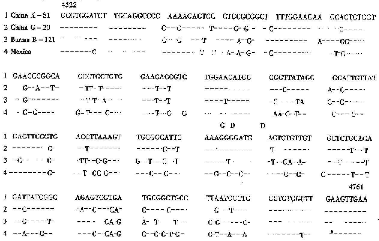
The amplified PCR positive products obtained from X-S1 and X-S6 samples were purified and ligated into pGEM-T vector, then transformed to E.coli JM 109. One of two white specks obtained from X-S1 had a band of about 239 bp in gel electrophoresis. Two white specks of the X-S6 were positive and a band of 239 bp was found after digested by EcoR I and Hind III. One positi ve clone of X-S1 was sequenced. HEV sequence of the X-S1 isolate could be reco gnized, this HEV X-S1 strain was neither lost nor inserted in length 239 bp compared with Burma strain of HEV. Aberrance occurred in 48 bases, 5 of them took place at the first codon position, 4 at the second position and 39 at the third codon position. The nucleotide sequence of X-S1 isolate is shown in Figure 1.
The sequence of HEV X-S1 strain was compared with the Chinese (G-20), Burmese (B-121) and Mexican strains of HEV. The similarity of nucleotide sequences was 85.4%, 79.2% and 76.4% respectively. The divergence of nucleotide sequences was 14.2%, 19.9% and 22.3% respectively.
This paper reports the results of serological survey on hepatitis E virus infection in Xiamen population. The serum samples from 81 patients with acute hepatitis were tested for anti-HEV IgG and IgM antibodies with HEV recombination antigen by ELISA. Twelve (14.8%) of 81 patients with acute hepatitis had antibody to HEV, 11 were positive for IgM anti-HEV and 2 positive for HEV RNA. The results show that there is hepatitis E virus infection in Xiamen again, but there has been no documented report of detection of HEV RNA in anti-HEV positive patients from this area.
The mixed infection of HEV and HBV were described previously[4]. This paper reports the co-infection of HEV and HAV. This case is positive for both ant ibodies of HAV and HEV. The patient presented as severe hepatitis in the clinica l characteristics, with very high ALT (20420 nmol·s-1/L). This shows that both the HEV and HAV are transmitted primarily through a fecal-oral route. In this study, the partial nucleotide sequence of Xiamen X-S1 isolate of sporad ic HEV was described and compared. It is shown that Xiamen (X-S1) strain and Guangzhou (G-20) strain[4] are most identical to each other (85.4%), wit h a lower range of identities to the Burmese strain[2] and Mexican strain[3] (80.1%-77.3%). The nucleotide sequences of the X-S1 strain and the G-20 strain may belong to a novel and unique branch. Similar results have been reported by other investigators[6,7]. Recently the HEV-US-1 strain was discovered by George G. Schlauder[8], it is significantly divergent f rom other human HEV isolates, which may be the fourth genotype. The discovery of these HEV variants may be important in understanding the worldwide distribution of HEV infection.
Edited by Jing-Yun Ma
| 1. | Gust ID, Purcell RH. Report of a workshop: waterborne non-A, non-B hepatitis. J Infect Dis. 1987;156:630-635. [PubMed] [DOI] [Cited in This Article: ] [Cited by in Crossref: 43] [Cited by in F6Publishing: 42] [Article Influence: 1.1] [Reference Citation Analysis (0)] |
| 2. | Tam AW, Smith MM, Guerra ME, Huang CC, Bradley DW, Fry KE, Reyes GR. Hepatitis E virus (HEV): molecular cloning and sequencing of the full-length viral genome. Virology. 1991;185:120-131. [PubMed] [DOI] [Cited in This Article: ] [Cited by in Crossref: 837] [Cited by in F6Publishing: 782] [Article Influence: 23.7] [Reference Citation Analysis (0)] |
| 3. | Huang CC, Nguyen D, Fernandez J, Yun KY, Fry KE, Bradley DW, Tam AW, Reyes GR. Molecular cloning and sequencing of the Mexico isolate of hepatitis E virus (HEV). Virology. 1992;191:550-558. [PubMed] [DOI] [Cited in This Article: ] [Cited by in Crossref: 261] [Cited by in F6Publishing: 264] [Article Influence: 8.3] [Reference Citation Analysis (0)] |
| 4. | Huang R, Nakazono N, Ishii K, Kawamata O, Kawaguchi R, Tsukada Y. Existing variations on the gene structure of hepatitis E virus strains from some regions of China. J Med Virol. 1995;47:303-308. [PubMed] [DOI] [Cited in This Article: ] [Cited by in Crossref: 55] [Cited by in F6Publishing: 58] [Article Influence: 2.0] [Reference Citation Analysis (0)] |
| 5. | Tsarev SA, Emerson SU, Reyes GR, Tsareva TS, Legters LJ, Malik IA, Iqbal M, Purcell RH. Characterization of a prototype strain of hepatitis E virus. Proc Natl Acad Sci USA. 1992;89:559-563. [PubMed] [DOI] [Cited in This Article: ] [Cited by in Crossref: 206] [Cited by in F6Publishing: 210] [Article Influence: 6.6] [Reference Citation Analysis (0)] |
| 6. | Wei SJ, Walsh P, Tong YB, Dong HQ, Cai XL. Nucleic acid se-quence analysis of the sporadic hepatitis E virus strains in Guangzhou. Chin J Microbiol Immunol. 1998;18:92-95. [Cited in This Article: ] |
| 7. | Wu JC, Sheen IJ, Chiang TY, Sheng WY, Wang YJ, Chan CY, Lee SD. The impact of traveling to endemic areas on the spread of hepatitis E virus infection: epidemiological and molecular analyses. Hepatology. 1998;27:1415-1420. [PubMed] [Cited in This Article: ] |
| 8. | Schlauder GG, Dawson GJ, Erker JC, Kwo PY, Knigge MF, Smalley DL, Rosenblatt JE, Desai SM, Mushahwar IK. The sequence and phylogenetic analysis of a novel hepatitis E virus isolated from a patient with acute hepatitis reported in the United States. J Gen Virol. 1998;79:447-456. [PubMed] [DOI] [Cited in This Article: ] [Cited by in Crossref: 250] [Cited by in F6Publishing: 251] [Article Influence: 9.7] [Reference Citation Analysis (0)] |


