Review Open Access
Copyright ©2009 The WJG Press and Baishideng. All rights reserved.
World J Gastroenterol. Jul 21, 2009; 15(27): 3349-3354
Published online Jul 21, 2009. doi: 10.3748/wjg.15.3349
Vitamin D for the prevention and treatment of pancreatic cancer
Kun-Chun Chiang, Department of Surgery, Chang Gung Memorial Hospital, Chang Gung University, Taoyuan 333, Taiwan, China
Tai C Chen, Boston University School of Medicine, 715 Albany Street, Boston, MA 02118, United States
Author contributions: Chiang KC wrote this manuscript; Chen TC revised the manuscript.
Correspondence to: Tai C Chen, Professor, Boston University School of Medicine, 715 Albany Street, Rm M-1022, Boston, MA 02118, United States. taichen@bu.edu
Telephone: +1-617-6384543
Fax: +1-617-6388898
Received: April 9, 2009
Revised: June 3, 2009
Accepted: June 10, 2009
Published online: July 21, 2009

Abstract

Pancreatic cancer is ranked fifth among cancer-related deaths worldwide with a 5-year survival rate of less than 5%. Currently, surgery is the only effective therapy. However, most patients are diagnosed in the late stage and are not suitable for receiving curative surgery. Moreover, pancreatic cancer doesn’t respond well to traditional chemotherapy and radiotherapy, leaving little effective treatment for advanced pancreatic cancer cases. 1α,25-dihydroxyvitamin D3 [1α,25(OH)2D3], the biologically active form of vitamin D3, was originally identified during studies of calcium and bone metabolism, though it is now recognized that it exerts biological effects in almost every tissue in the body. Abundant evidence has shown that 1α,25(OH)2D3 has antiproliferative, apoptotic, pro-differentiation and antiangiogensis effects in many types of cancer cells in vivo and in vitro, including breast, prostate, and colon. Similarly, the antitumor growth effect of 1α,25(OH)2D3 on pancreatic cells has been demonstrated. The clinical use of 1α,25(OH)2D3 is impeded by the lethal side effects of hypercalcemia and hypercalciuria. Therefore, 1α,25(OH)2D3 analogs, which are either equipotent or more potent than 1α,25(OH)2D3 in inhibiting tumor cell growth but with fewer hypercalcemic and hypercalciuric side effects, have been developed for the treatment of different cancers. Recently, a pre-clinical study demonstrated that a less calcemic analog of 1α,25(OH)2D3, 19-nor-1α,25(OH)2D2 (Paricalcitol), is effective in inhibiting tumor growth in vitro and in vivo, via upregulation of p21 and p27 tumor suppressor genes. Studies on the anti-tumor effects of a more potent analog of Paricalcitol are underway. 1α,25(OH)2D3 and its analogs are potentially attractive novel therapies for pancreatic cancer.

Key Words: Vitamin D, Pancreatic cancer, Calcitriol, Paricalcitol, Chemoprevention, Adenocarcinoma, CYP27b1


INTRODUCTION

Pancreatic adenocarcinoma is one of the most lethal human malignancies. It ranks as the fourth most common cancer-related mortality in the Western world and fifth most common worldwide[1]. There were 37 680 new cases of pancreatic cancer and 34 290 deaths due to this disease in 2008 in the Unites States alone[2]. The overall 5-year survival rate of pancreatic cancer is estimated to be around 1%-4%, which is attributed to its aggressive growth behavior, such as early local spread and metastasis, and resistance to radiation and most systemic chemotherapies[1]. The poor prognosis is also due to the lack of effective early diagnosis. Consequently, most patients have been diagnosed with late stage disease. Currently, surgery remains the cornerstone of treatment. After resection, the 5-year survival rate is 10%-29%[35], but only 10%-15% of pancreatic cancer patients are suitable candidates for resection. For the non-operable pancreatic cancer cases, the most frequently used treatment methods include radiation and chemotherapy with 5-fluorouracil, cisplatin or gemcitabine[68], or a combination of these modalities[911]. Growing evidence supports neoadjuvant therapy to downstage some pancreatic cancer patients from borderline resectable to resectable disease[1213], but its impact on long-term survival still needs further examination[14]. With this bleak background, there is an urgent need to develop novel treatments for pancreatic cancer.

SOURCES AND METABOLISM OF VITAMIN D

The production of vitamin D3 depends on solar ultraviolet B radiation (wavelength between 290-315 nm), which converts 7-dehydrocholesterol stored in the skin to previtamin D3. Previtamin D3 is then thermoisomerized to vitamin D3, which then enters the bloodstream and is bound to the vitamin D binding protein. Very little food naturally contains vitamin D. The cutaneous synthesis of vitamin D3 is normally responsible for over 90% of our vitamin D requirement[15]. Vitamin D (including both vitamin D2 and vitamin D3) obtained from sunlight or dietary sources is then converted to 25-hydroxyvitamin D [25(OH)D], catalyzed by vitamin D-25-hydroxylase in the liver. 25(OH)D is the major circulating form of vitamin D and is widely accepted as an index of vitamin D status in humans. However, it is biologically inert until it is hydroxylated in the kidney to form 1α,25-dihydroxyvitamin D [1α,25(OH)2D]. 1α,25(OH)2D is a lipid-soluble hormone that interacts with the vitamin D receptor (VDR) to exert a variety of functions, including genomic and non-genomic actions.

VITAMIN D AND CANCER

1α,25(OH)2D, the biologically active form of vitamin D, was originally discovered because of its effects on calcium and bone metabolism. It is now recognized that the hormone has activities in almost every tissue in the body. 1α,25(OH)2D3 implements this effect through binding to the nuclear VDR and then binding to a specific DNA sequence, the vitamin D response elements (VDREs)[16]. Via this genomic pathway, 1α,25(OH)2D3 can modulate gene expression in a tissue-specific manner, mainly leading to inhibition of cellular proliferation, induction of differentiation and apoptosis, which in turn, protect cells from malignant transformation and repress cancer cell growth.

However, the use of vitamin D to treat cancers has been impeded by lethal hypercalcemia induced by systemic administration of 1α,25(OH)2D3. To overcome this drawback, analogs of 1α,25(OH)2D3, exhibiting more potent growth inhibition but less calcemic effects, have been developed as anticancer drugs[17], and some of them have shown great results in pre-clinical studies. For example, Akhter et al[18] demonstrated that a 1α,25(OH)2D3 analog, EB 1089, exhibited an antiproliferative effect on colon cancer in a xenograft animal model. Abe-Hashimoto et al[19] showed that another 1α,25(OH)2D3 analog, OCT, displayed an antitumor effect on a xenograft model using MCF-7, a breast cancer cell line, in combination with tamoxifen. Furthermore, Polek et al[20] showed that another analog, LG190119, possessed antiproliferative activity when tested in an LNCaP prostate cancer cell xenograft model.

There are some limited clinical trials that show 1α,25(OH)2D3 and its analogs have antitumor effects in humans when used alone or in combination with other chemotherapy drugs. For example, Beer et al[21] conducted a phase II clinical trial in which they showed the combination of 1α,25(OH)2D3 and docetaxel could induce > 50% decline in PSA, a prostate cancer tumor marker, and improve survival of prostate cancer patients. However, most of the clinical trials only confirmed the less- or non-calcemic effect of 1α,25(OH)2D3 or its analog when used alone or in combination with other chemotherapy drugs without prolonging the survival of cancer patients. There are still many ongoing clinical trials attempting to demonstrate the antitumor effect of 1α,25(OH)2D3 and its analogs in humans. More effort is required in order to fully appreciate the clinical utilities of vitamin D-based therapies in treating human diseases, including cancers.

MECHANISMS OF VITAMIN D ACTIONS FOR CANCER TREATMENT

Vitamin D reduces the risk of cancer through its biologically active metabolite, 1α,25(OH)2D3, which regulates cellular proliferation and differentiation, inhibits angiogenesis, and induces apoptosis.

Stumpf et al[22], in 1979, reported that the VDR existed not only in the intestine, bone and kidney, but in almost all tissues in the body. Suda et al[23] (1982) first noted that 1α,25(OH)2D3 caused a marked inhibition of cell growth on VDR- positive M-1 leukemic cells. In the same year, Tanaka et al[24] reported that 1α,25(OH)2D3 showed the same effect on HL-60 leukemic cells, which had the VDR. Since then, many VDR-containing cancer cell lines, including prostate, colon, breast, lung, and melanoma, have shown growth inhibition when exposed to 1α,25(OH)2D3[152527].

The antiproliferative effects of 1α,25(OH)2D3 are mainly due to alterations in several key regulators of the cell cycle, culminating in dephosphorylation of retinoblastoma protein and arrest of cells in G0/G1[28]. Progression of the cell cycle is regulated by cyclins and their associated cyclin dependant kinases and cyclin dependant kinase inhibitors (CKIs). The CKI genes, such as p21 and/or p27, have VDRE within their promoter regions, and are genomic targets of the 1α,25(OH)2D3/VDR complex in many cell types, which in turn, induces G1 cell-cycle arrest and withdrawal from the cell cycle[2931]. However, some genes are transcriptionally affected by 1α,25(OH)2D3 but lack VDREs in their promoter regions, which suggests that 1α,25(OH)2D3 induces indirect effects on cell-cycle regulation through another signaling pathway. For example, 1α,25(OH)2D3 could downregulate the expression of estrogen, epidermal growth factor, insulin-like growth factor 1, and keratinocyte growth factor and upregulate inhibitory growth factors, such as transformation growth factor-β[3235].

1α,25(OH)2D3 can also induce differentiation to control tumor cell proliferation through the pathways of phosphatidylinositol 3-kinase, which is VDR dependent[36], and by suppression of interleukin 12 protein secretion[37], which is VDR independent.

Induction of apoptosis is another important function of 1α,25(OH)2D3, which represses the expression of the antiapoptotic protein, BCL2, and prosurvival protein, BCL-XL. 1α,25(OH)2D3 also enhances the expression of proapoptotic proteins such as BAX and BAD[38]. In addition to the BCL2 family, 1α,25(OH)2D3 can activate the caspase effector molecules directly to induce apoptosis[38]. Furthermore, it has been shown that 1α,25(OH)2D3, in combination with radiation or chemotherapeutic agents, causes an additive effect on cancer cell death[3942].

Inhibition of angiogenesis is also an important anticancer mechanism of 1α,25(OH)2D3. 1α,25(OH)2D3 can repress endothelial cell growth in vitro and reduce angiogenesis in vivo[4345]. The anti-angiogenic effects of 1α,25(OH)2D3 might subsequently lead to the inhibition of metastasis, as demonstrated in murine prostate and lung models treated with 1α,25(OH)2D3[4647].

The precise anticancer mechanisms of 1α,25(OH)2D3, including VDR-dependent and VDR-independent pathways are not fully understood. For the further use of 1α,25(OH)2D3 and its analogs in the treatment of cancers, there should be more studies addressing this issue to pave the way for the development of more potent 1α,25(OH)2D3 analogs.

ASSOCIATION OF VITAMIN D AND PANCREATIC CANCER-EPIDEMIOLOGICAL EVIDENCE

Epidemiologic studies have shown that vitamin D status, influenced by living at high or low latitude, solar UV exposure and dietary intake of vitamin D, was inversely associated with the incidence of some cancers such as prostate, colon and breast[4850]. It was also reported that the risk of developing prostate, breast and colon cancer was decreased by 30%-50% if the serum concentration of 25(OH)D exceeded 50 nmol/L[5152]. Tangpricha et al[53] demonstrated that vitamin D deficiency enhanced the growth of MC-26 colon cancer xenografts in BALB/c mice, which supported the hypothesis that vitamin D sufficiency could reduce the proliferation of tumor cells in vivo.

To date, there have been only two epidemiological studies discussing the vitamin D status and the incidence of pancreatic cancer. One was conducted by Skinner et al[54] on U.S. nurses and health professionals, which demonstrated that higher dietary intake of vitamin D could be associated with lower incidence of pancreatic cancer. The other report, however, conducted in the observational cohort of the Finnish Alpha-Tocopheral Beta-Carotene (ATBC) trial, showed a discrepancy in results compared to the report by Skinner et al[54]. The ATBC data suggested that a higher serum concentration of 25(OH)D in male smokers was associated with a higher incidence of pancreatic cancer[55]. The reason behind these contradictory results was not clear, but they employed different measures of dietary intake of vitamin D versus 25(OH)D, and the participants in the ATBC trials were all male smokers, which might have contributed to the different conclusions in the two studies. To clarify this, more studies addressing the relationship between vitamin D status and incidence of pancreatic cancer are required.

VITAMIN D AND PANCREATIC CANCER-BIOCHEMICAL EVIDENCE

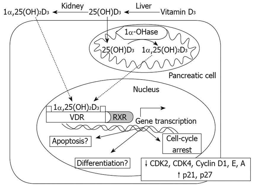
While the precise anticancer mechanisms, including VDR-dependent and VDR-independent actions, induced by 1α,25(OH)2D3 still require further investigation, it is known that in pancreatic cancer cells, 1α,25(OH)2D3 induces the expression of p21 and p27 and inhibits the production of cyclins (D1, E and A) and cyclin dependent kinases 2 and 4, which in turn, elicit cell cycle arrest in G0/G1in vitro[56]. Similar effects have been observed with 19-nor-1α,25(OH)2D2 (Paricalcitol) in human pancreatic cancer cells in vitro and in vivo by upregulating the expressions of p21 and p27[57] (Figure 1).

Figure 1
Figure 1 Mechanisms of Vitamin D3 action on pancreas cells. Both 1,25(OH)2D3 and 25(OH)D3 can enter pancreas cells, where 25(OH)D3 can be converted to 1,25(OH)2D3 by 1α-OHase. 1,25(OH)2D3 can bind the VDR and further combine with the retinoid X receptor (RXR) to form a heterodimer, VDR-RXR. The VDR-RXR heterodimer binds to specific vitamin D response elements located in the promoter region of vitamin D-responsive genes, which in turn induces gene transcription. So far, in terms of pancreas cells, inhibition of CDK2, CDK4, Cyclin D1, Cyclin E, Cyclin A and upregulation of p21 and p27 have been demonstrated, leading to a block in the cell cycle at G0/G1. Regarding induction of differentiation, apoptosis and other anticancer mechanisms, further studies are required.

Currently, very little is known about the mechanisms of vitamin D actions on pancreatic cancer cells. More emphasis in this area is needed, because very few efficient therapeutic options are available to benefit pancreatic cancer survival.

POTENTIAL OF VITAMIN D ANALOGS FOR PANCREATIC CANCER TREATMENT

Since the VDR exists in almost every tissue of the body[22] and 1α,25(OH)2D3 exhibits growth inhibitory effects on may different types of cancer cells, several studies have been performed to investigate the anticancer effect of vitamin D and its analogs on pancreatic cancer cells during the past two decades. For example, Kawa et al[56] reported that 22-oxa-1α,25(OH)2D3, a 1α,25(OH)2D3 analog, caused growth inhibition in three pancreatic cancer cell lines and inhibited the growth of a BxPC-3 tumor in a xenograft nude mice model[58]. Colston et al[59] demonstrated that another 1α,25(OH)2D3 analog, EB 1089, exhibited more potent antitumor effects than 1α,25(OH)2D3 on the GER cell line, a human pancreatic cancer cell line, in vitro and in vivo. Pettersson et al[60] also reported that EB 1089 induced greater tumor growth inhibition than 9-cis-retinoic acid in vitro. These two pre-clinical studies therefore suggested that EB 1089 might be a promising vitamin D analog for further clinical trials in pancreatic cancer patients. However, in a phase II clinical trial, which is also the only clinical trial studying the use of vitamin D analogs against pancreatic cancer, EB 1089 given once daily orally failed to significantly improve patient survival, although most patients tolerated the daily orally dose of 10-15 &mgr;g of the drug well, without causing hypercalcemia[61].

With the knowledge that 19-nor-1α,25(OH)2D2 (Paricalcitol) is a Federal Drug Administration approved drug for the treatment and prevention of secondary hyperparathyroidism associated with chronic kidney disease[62], and this analog has comparable growth inhibitory effects as 1α,25(OH)2D3 in human prostate cancer cells in vitro[63], Schwartz et al[57] studied the same analog in human pancreatic cancer cells in vitro and demonstrated that it also inhibited the proliferation of these cells. Given the fact that Paricalcitol has been shown to be less calcemic than 1α,25(OH)2D and few therapeutic options for pancreatic cancer patients are available, it is worth further exploration as an anticancer drug for pancreatic cancer.

In addition to using synthetic 1α,25(OH)2D3 analogs to treat pancreatic cancer in an effort to avoid hypercalcemia, another strategy is the employment of inactive prohormone 25(OH)D3 (Figure 1). This is based on the finding that many normal and cancerous cells, including prostate cell lines and primary cultures of prostate cells, possess 25(OH)D-1α-hydroxylase (1α-hydroxylase or CYP27b1), the enzyme responsible for the conversion of 25(OH)D3 to 1α,25(OH)2D3[26], and CYP27b1 has been proposed as a tumor suppressor[64]. Furthermore, it has been demonstrated that 25(OH)D3 had a comparable tumor growth inhibitory effect to 1α,25(OH)2D3 on primary cultures of prostate cells[6365]. These findings led to a subsequent study demonstrating that CYP27b1 activity was also expressed in pancreatic cancer cells and the growth of these cancer cells was inhibited in the presence of 25(OH)D3[66]. Given the fact that the conversion of 25(OH)D3 to 1α,25(OH)2D3 occurs within the cell, and the 1α,25(OH)2D3 formed acts in a autocrine/paracrine fashion[266365], systemic administration of 25(OH)D3 might greatly minimize the risk of hypercalcemia. 25(OH)D3 has been approved by the Federal Drug Administration to treat vitamin D deficiency in humans; therefore, 25(OH)D3 could be another attractive candidate for clinical trials in pancreatic cancer patients.

CONCLUSION

Abundant evidence indicates that 1α,25(OH)2D3 has antiproliferative, pro-differentiative, apoptotic and antiangiogenic activities in different types of tumor cells. In different cancer cell types, 1α,25(OH)2D3 can induce the expression of various molecular markers to regulate cell growth. Even in the same tissue, different cell types might exhibit heterogeneous responses to the addition of 1α,25(OH)2D3. It is known that 1α,25(OH)2D3 and its analogs can inhibit pancreatic cancer cell growth in vitro[5659] via activation of p21 and p27, which in turn influence cell cycle progression and arrest cells in G0/G1. So far, the only pancreatic cancer clinical trial using a 1α,25(OH)2D3 analog, EB 1089[61], did not show significant prolongation of survival. However, a preclinical study with 19-nor-1α,25(OH)2D2 (Paricalcitol) in pancreatic cancer cells[60] has provided some encouraging results and might lead to further clinical trials. Moreover, given the fact that pancreatic cancer cells posses 1α-hydroxylase (CYP27b1) activity and can convert the prohormone 25(OH)D, which has a low risk of inducing hypercalcemia even at high concentrations, into 1α,25(OH)2D within the cells and thereby inhibiting pancreatic cancer cell growth in a autocrine/paracrine fashion[266365], prohormone 25(OH)D-based therapy might be another attractive treatment strategy for pancreatic cancer in the future.

Footnotes

Peer reviewers: Dario Conte, Professor, GI Unit - IRCCS Osp. Maggiore, Chair of Gastroenterology, Via F. Sforza, 35, Milano 20122, Italy; Ian C Roberts-Thomson, Professor, Department of Gastroenterology and Hepatology, The Queen Elizabeth Hospital, 28 Woodville Road, Woodville South 5011, Australia

References
1.  Jemal A, Siegel R, Ward E, Murray T, Xu J, Thun MJ. Cancer statistics, 2007. CA Cancer J Clin. 2007;57:43-66.  [PubMed]  [DOI]  [Cited in This Article: ]
2.  American Cancer Society Cancer Facts and Figures 2008. Atlanta: American Cancer society 2008;  Available from: URL: http://www.cancer.org/downloads/STT/2008CAFFfinalsecured.pdf.  [PubMed]  [DOI]  [Cited in This Article: ]
3.  Trede M, Schwall G, Saeger HD. Survival after pancreatoduodenectomy. 118 consecutive resections without an operative mortality. Ann Surg. 1990;211:447-458.  [PubMed]  [DOI]  [Cited in This Article: ]
4.  Yeo CJ, Cameron JL, Sohn TA, Lillemoe KD, Pitt HA, Talamini MA, Hruban RH, Ord SE, Sauter PK, Coleman J. Six hundred fifty consecutive pancreaticoduodenectomies in the 1990s: pathology, complications, and outcomes. Ann Surg. 1997;226:248-257; discussion 257-260.  [PubMed]  [DOI]  [Cited in This Article: ]
5.  Nitecki SS, Sarr MG, Colby TV, van Heerden JA. Long-term survival after resection for ductal adenocarcinoma of the pancreas. Is it really improving? Ann Surg. 1995;221:59-66.  [PubMed]  [DOI]  [Cited in This Article: ]
6.  Heinemann V. Gemcitabine: progress in the treatment of pancreatic cancer. Oncology. 2001;60:8-18.  [PubMed]  [DOI]  [Cited in This Article: ]
7.  Rocha Lima CM, Urbanic JJ, Lal A, Kneuper-Hall R, Brunson CY, Green MR. Beyond pancreatic cancer: irinotecan and gemcitabine in solid tumors and hematologic malignancies. Semin Oncol. 2001;28:34-43.  [PubMed]  [DOI]  [Cited in This Article: ]
8.  Ward S, Morris E, Bansback N, Calvert N, Crellin A, Forman D, Larvin M, Radstone D. A rapid and systematic review of the clinical effectiveness and cost-effectiveness of gemcitabine for the treatment of pancreatic cancer. Health Technol Assess. 2001;5:1-70.  [PubMed]  [DOI]  [Cited in This Article: ]
9.  Stathopoulos GP, Mavroudis D, Tsavaris N, Kouroussis C, Aravantinos G, Agelaki S, Kakolyris S, Rigatos SK, Karabekios S, Georgoulias V. Treatment of pancreatic cancer with a combination of docetaxel, gemcitabine and granulocyte colony-stimulating factor: a phase II study of the Greek Cooperative Group for Pancreatic Cancer. Ann Oncol. 2001;12:101-103.  [PubMed]  [DOI]  [Cited in This Article: ]
10.  Crane CH, Wolff RA, Abbruzzese JL, Evans DB, Milas L, Mason K, Charnsangavej C, Pisters PW, Lee JE, Lenzi R. Combining gemcitabine with radiation in pancreatic cancer: understanding important variables influencing the therapeutic index. Semin Oncol. 2001;28:25-33.  [PubMed]  [DOI]  [Cited in This Article: ]
11.  Kozuch P, Grossbard ML, Barzdins A, Araneo M, Robin A, Frager D, Homel P, Marino J, DeGregorio P, Bruckner HW. Irinotecan combined with gemcitabine, 5-fluorouracil, leucovorin, and cisplatin (G-FLIP) is an effective and noncrossresistant treatment for chemotherapy refractory metastatic pancreatic cancer. Oncologist. 2001;6:488-495.  [PubMed]  [DOI]  [Cited in This Article: ]
12.  Massucco P, Capussotti L, Magnino A, Sperti E, Gatti M, Muratore A, Sgotto E, Gabriele P, Aglietta M. Pancreatic resections after chemoradiotherapy for locally advanced ductal adenocarcinoma: analysis of perioperative outcome and survival. Ann Surg Oncol. 2006;13:1201-1208.  [PubMed]  [DOI]  [Cited in This Article: ]
13.  Greer SE, Pipas JM, Sutton JE, Zaki BI, Tsapakos M, Colacchio TA, Gibson JJ, Wiener DC, Ripple GH, Barth RJ Jr. Effect of neoadjuvant therapy on local recurrence after resection of pancreatic adenocarcinoma. J Am Coll Surg. 2008;206:451-457.  [PubMed]  [DOI]  [Cited in This Article: ]
14.  van Moorsel CJ, Veerman G, Bergman AM, Guechev A, Vermorken JB, Postmus PE, Peters GJ. Combination chemotherapy studies with gemcitabine. Semin Oncol. 1997;24:S7-17-S7-23.  [PubMed]  [DOI]  [Cited in This Article: ]
15.  Holick MF. Sunlight and vitamin D for bone health and prevention of autoimmune diseases, cancers, and cardiovascular disease. Am J Clin Nutr. 2004;80:1678S-1688S.  [PubMed]  [DOI]  [Cited in This Article: ]
16.  Tsai MJ, O'Malley BW. Molecular mechanisms of action of steroid/thyroid receptor superfamily members. Annu Rev Biochem. 1994;63:451-486.  [PubMed]  [DOI]  [Cited in This Article: ]
17.  Guyton KZ, Kensler TW, Posner GH. Cancer chemoprevention using natural vitamin D and synthetic analogs. Annu Rev Pharmacol Toxicol. 2001;41:421-442.  [PubMed]  [DOI]  [Cited in This Article: ]
18.  Akhter J, Chen X, Bowrey P, Bolton EJ, Morris DL. Vitamin D3 analog, EB1089, inhibits growth of subcutaneous xenografts of the human colon cancer cell line, LoVo, in a nude mouse model. Dis Colon Rectum. 1997;40:317-321.  [PubMed]  [DOI]  [Cited in This Article: ]
19.  Abe-Hashimoto J, Kikuchi T, Matsumoto T, Nishii Y, Ogata E, Ikeda K. Antitumor effect of 22-oxa-calcitriol, a noncalcemic analogue of calcitriol, in athymic mice implanted with human breast carcinoma and its synergism with tamoxifen. Cancer Res. 1993;53:2534-2537.  [PubMed]  [DOI]  [Cited in This Article: ]
20.  Polek TC, Murthy S, Blutt SE, Boehm MF, Zou A, Weigel NL, Allegretto EA. Novel nonsecosteroidal vitamin D receptor modulator inhibits the growth of LNCaP xenograft tumors in athymic mice without increased serum calcium. Prostate. 2001;49:224-233.  [PubMed]  [DOI]  [Cited in This Article: ]
21.  Beer TM, Ryan CW, Venner PM, Petrylak DP, Chatta GS, Ruether JD, Redfern CH, Fehrenbacher L, Saleh MN, Waterhouse DM. Double-blinded randomized study of high-dose calcitriol plus docetaxel compared with placebo plus docetaxel in androgen-independent prostate cancer: a report from the ASCENT Investigators. J Clin Oncol. 2007;25:669-674.  [PubMed]  [DOI]  [Cited in This Article: ]
22.  Stumpf WE, Sar M, Reid FA, Tanaka Y, DeLuca HF. Target cells for 1,25-dihydroxyvitamin D3 in intestinal tract, stomach, kidney, skin, pituitary, and parathyroid. Science. 1979;206:1188-1190.  [PubMed]  [DOI]  [Cited in This Article: ]
23.  Suda T, Abe E. Induction of differentiation of human myeloid leukemia cells by 1,25-dihydroxyvitamin D3. Vitamin D: chemical, biochemical and clinical endocrinology of calcium metabolism. Walter de Gruyter: New York 1982; 59-64.  [PubMed]  [DOI]  [Cited in This Article: ]
24.  Tanaka H, Abe E, Miyaura C, Kuribayashi T, Konno K, Nishii Y, Suda T. 1 alpha,25-Dihydroxycholecalciferol and a human myeloid leukaemia cell line (HL-60). Biochem J. 1982;204:713-719.  [PubMed]  [DOI]  [Cited in This Article: ]
25.  Colston K, Hirt M, Feldman D. Organ distribution of the cytoplasmic 1,25-dihydroxycholecalciferol receptor in various mouse tissues. Endocrinology. 1980;107:1916-1922.  [PubMed]  [DOI]  [Cited in This Article: ]
26.  Chen TC, Holick MF. Vitamin D and prostate cancer prevention and treatment. Trends Endocrinol Metab. 2003;14:423-430.  [PubMed]  [DOI]  [Cited in This Article: ]
27.  Skowronski RJ, Peehl DM, Feldman D. Vitamin D and prostate cancer: 1,25 dihydroxyvitamin D3 receptors and actions in human prostate cancer cell lines. Endocrinology. 1993;132:1952-1960.  [PubMed]  [DOI]  [Cited in This Article: ]
28.  Simboli-Campbell M, Narvaez CJ, van Weelden K, Tenniswood M, Welsh J. Comparative effects of 1,25(OH)2D3 and EB1089 on cell cycle kinetics and apoptosis in MCF-7 breast cancer cells. Breast Cancer Res Treat. 1997;42:31-41.  [PubMed]  [DOI]  [Cited in This Article: ]
29.  Inoue T, Kamiyama J, Sakai T. Sp1 and NF-Y synergistically mediate the effect of vitamin D(3) in the p27(Kip1) gene promoter that lacks vitamin D response elements. J Biol Chem. 1999;274:32309-32317.  [PubMed]  [DOI]  [Cited in This Article: ]
30.  Hager G, Formanek M, Gedlicka C, Thurnher D, Knerer B, Kornfehl J. 1,25(OH)2 vitamin D3 induces elevated expression of the cell cycle-regulating genes P21 and P27 in squamous carcinoma cell lines of the head and neck. Acta Otolaryngol. 2001;121:103-109.  [PubMed]  [DOI]  [Cited in This Article: ]
31.  Wu G, Fan RS, Li W, Ko TC, Brattain MG. Modulation of cell cycle control by vitamin D3 and its analogue, EB1089, in human breast cancer cells. Oncogene. 1997;15:1555-1563.  [PubMed]  [DOI]  [Cited in This Article: ]
32.  Cordero JB, Cozzolino M, Lu Y, Vidal M, Slatopolsky E, Stahl PD, Barbieri MA, Dusso A. 1,25-Dihydroxyvitamin D down-regulates cell membrane growth- and nuclear growth-promoting signals by the epidermal growth factor receptor. J Biol Chem. 2002;277:38965-38971.  [PubMed]  [DOI]  [Cited in This Article: ]
33.  Kawata H, Kamiakito T, Takayashiki N, Tanaka A. Vitamin D3 suppresses the androgen-stimulated growth of mouse mammary carcinoma SC-3 cells by transcriptional repression of fibroblast growth factor 8. J Cell Physiol. 2006;207:793-799.  [PubMed]  [DOI]  [Cited in This Article: ]
34.  Wu Y, Craig TA, Lutz WH, Kumar R. Identification of 1 alpha,25-dihydroxyvitamin D3 response elements in the human transforming growth factor beta 2 gene. Biochemistry. 1999;38:2654-2660.  [PubMed]  [DOI]  [Cited in This Article: ]
35.  Xie SP, Pirianov G, Colston KW. Vitamin D analogues suppress IGF-I signalling and promote apoptosis in breast cancer cells. Eur J Cancer. 1999;35:1717-1723.  [PubMed]  [DOI]  [Cited in This Article: ]
36.  Hmama Z, Nandan D, Sly L, Knutson KL, Herrera-Velit P, Reiner NE. 1alpha,25-dihydroxyvitamin D(3)-induced myeloid cell differentiation is regulated by a vitamin D receptor-phosphatidylinositol 3-kinase signaling complex. J Exp Med. 1999;190:1583-1594.  [PubMed]  [DOI]  [Cited in This Article: ]
37.  Penna G, Adorini L. 1 Alpha,25-dihydroxyvitamin D3 inhibits differentiation, maturation, activation, and survival of dendritic cells leading to impaired alloreactive T cell activation. J Immunol. 2000;164:2405-2411.  [PubMed]  [DOI]  [Cited in This Article: ]
38.  Ylikomi T, Laaksi I, Lou YR, Martikainen P, Miettinen S, Pennanen P, Purmonen S, Syvälä H, Vienonen A, Tuohimaa P. Antiproliferative action of vitamin D. Vitam Horm. 2002;64:357-406.  [PubMed]  [DOI]  [Cited in This Article: ]
39.  Chaudhry M, Sundaram S, Gennings C, Carter H, Gewirtz DA. The vitamin D3 analog, ILX-23-7553, enhances the response to adriamycin and irradiation in MCF-7 breast tumor cells. Cancer Chemother Pharmacol. 2001;47:429-436.  [PubMed]  [DOI]  [Cited in This Article: ]
40.  Posner GH, Crawford KR, Peleg S, Welsh JE, Romu S, Gewirtz DA, Gupta MS, Dolan P, Kensler TW. A non-calcemic sulfone version of the vitamin D(3) analogue seocalcitol (EB 1089): chemical synthesis, biological evaluation and potency enhancement of the anticancer drug adriamycin. Bioorg Med Chem. 2001;9:2365-2671.  [PubMed]  [DOI]  [Cited in This Article: ]
41.  Sundaram S, Gewirtz DA. The vitamin D3 analog EB 1089 enhances the response of human breast tumor cells to radiation. Radiat Res. 1999;152:479-486.  [PubMed]  [DOI]  [Cited in This Article: ]
42.  Sundaram S, Sea A, Feldman S, Strawbridge R, Hoopes PJ, Demidenko E, Binderup L, Gewirtz DA. The combination of a potent vitamin D3 analog, EB 1089, with ionizing radiation reduces tumor growth and induces apoptosis of MCF-7 breast tumor xenografts in nude mice. Clin Cancer Res. 2003;9:2350-2356.  [PubMed]  [DOI]  [Cited in This Article: ]
43.  Chung I, Wong MK, Flynn G, Yu WD, Johnson CS, Trump DL. Differential antiproliferative effects of calcitriol on tumor-derived and matrigel-derived endothelial cells. Cancer Res. 2006;66:8565-8573.  [PubMed]  [DOI]  [Cited in This Article: ]
44.  Merke J, Milde P, Lewicka S, Hügel U, Klaus G, Mangelsdorf DJ, Haussler MR, Rauterberg EW, Ritz E. Identification and regulation of 1,25-dihydroxyvitamin D3 receptor activity and biosynthesis of 1,25-dihydroxyvitamin D3. Studies in cultured bovine aortic endothelial cells and human dermal capillaries. J Clin Invest. 1989;83:1903-1915.  [PubMed]  [DOI]  [Cited in This Article: ]
45.  Iseki K, Tatsuta M, Uehara H, Iishi H, Yano H, Sakai N, Ishiguro S. Inhibition of angiogenesis as a mechanism for inhibition by 1alpha-hydroxyvitamin D3 and 1,25-dihydroxyvitamin D3 of colon carcinogenesis induced by azoxymethane in Wistar rats. Int J Cancer. 1999;81:730-733.  [PubMed]  [DOI]  [Cited in This Article: ]
46.  Getzenberg RH, Light BW, Lapco PE, Konety BR, Nangia AK, Acierno JS, Dhir R, Shurin Z, Day RS, Trump DL. Vitamin D inhibition of prostate adenocarcinoma growth and metastasis in the Dunning rat prostate model system. Urology. 1997;50:999-1006.  [PubMed]  [DOI]  [Cited in This Article: ]
47.  Nakagawa K, Kawaura A, Kato S, Takeda E, Okano T. 1 alpha,25-Dihydroxyvitamin D(3) is a preventive factor in the metastasis of lung cancer. Carcinogenesis. 2005;26:429-440.  [PubMed]  [DOI]  [Cited in This Article: ]
48.  Garland CF, Garland FC. Do sunlight and vitamin D reduce the likelihood of colon cancer? Int J Epidemiol. 1980;9:227-231.  [PubMed]  [DOI]  [Cited in This Article: ]
49.  Schwartz GG, Hulka BS. Is vitamin D deficiency a risk factor for prostate cancer? (Hypothesis). Anticancer Res. 1990;10:1307-1311.  [PubMed]  [DOI]  [Cited in This Article: ]
50.  Gorham ED, Garland FC, Garland CF. Sunlight and breast cancer incidence in the USSR. Int J Epidemiol. 1990;19:820-824.  [PubMed]  [DOI]  [Cited in This Article: ]
51.  Ahonen MH, Tenkanen L, Teppo L, Hakama M, Tuohimaa P. Prostate cancer risk and prediagnostic serum 25-hydroxyvitamin D levels (Finland). Cancer Causes Control. 2000;11:847-852.  [PubMed]  [DOI]  [Cited in This Article: ]
52.  Garland CF, Comstock GW, Garland FC, Helsing KJ, Shaw EK, Gorham ED. Serum 25-hydroxyvitamin D and colon cancer: eight-year prospective study. Lancet. 1989;2:1176-1178.  [PubMed]  [DOI]  [Cited in This Article: ]
53.  Tangpricha V, Spina C, Yao M, Chen TC, Wolfe MM, Holick MF. Vitamin D deficiency enhances the growth of MC-26 colon cancer xenografts in Balb/c mice. J Nutr. 2005;135:2350-2354.  [PubMed]  [DOI]  [Cited in This Article: ]
54.  Skinner HG, Michaud DS, Giovannucci E, Willett WC, Colditz GA, Fuchs CS. Vitamin D intake and the risk for pancreatic cancer in two cohort studies. Cancer Epidemiol Biomarkers Prev. 2006;15:1688-1695.  [PubMed]  [DOI]  [Cited in This Article: ]
55.  Stolzenberg-Solomon RZ, Vieth R, Azad A, Pietinen P, Taylor PR, Virtamo J, Albanes D. A prospective nested case-control study of vitamin D status and pancreatic cancer risk in male smokers. Cancer Res. 2006;66:10213-10219.  [PubMed]  [DOI]  [Cited in This Article: ]
56.  Kawa S, Nikaido T, Aoki Y, Zhai Y, Kumagai T, Furihata K, Fujii S, Kiyosawa K. Vitamin D analogues up-regulate p21 and p27 during growth inhibition of pancreatic cancer cell lines. Br J Cancer. 1997;76:884-889.  [PubMed]  [DOI]  [Cited in This Article: ]
57.  Schwartz GG, Eads D, Naczki C, Northrup S, Chen T, Koumenis C. 19-nor-1 alpha,25-dihydroxyvitamin D2 (paricalcitol) inhibits the proliferation of human pancreatic cancer cells in vitro and in vivo. Cancer Biol Ther. 2008;7:430-436.  [PubMed]  [DOI]  [Cited in This Article: ]
58.  Kawa S, Yoshizawa K, Tokoo M, Imai H, Oguchi H, Kiyosawa K, Homma T, Nikaido T, Furihata K. Inhibitory effect of 220-oxa-1,25-dihydroxyvitamin D3 on the proliferation of pancreatic cancer cell lines. Gastroenterology. 1996;110:1605-1613.  [PubMed]  [DOI]  [Cited in This Article: ]
59.  Colston KW, James SY, Ofori-Kuragu EA, Binderup L, Grant AG. Vitamin D receptors and anti-proliferative effects of vitamin D derivatives in human pancreatic carcinoma cells in vivo and in vitro. Br J Cancer. 1997;76:1017-1020.  [PubMed]  [DOI]  [Cited in This Article: ]
60.  Pettersson F, Colston KW, Dalgleish AG. Differential and antagonistic effects of 9-cis-retinoic acid and vitamin D analogues on pancreatic cancer cells in vitro. Br J Cancer. 2000;83:239-245.  [PubMed]  [DOI]  [Cited in This Article: ]
61.  Evans TR, Colston KW, Lofts FJ, Cunningham D, Anthoney DA, Gogas H, de Bono JS, Hamberg KJ, Skov T, Mansi JL. A phase II trial of the vitamin D analogue Seocalcitol (EB1089) in patients with inoperable pancreatic cancer. Br J Cancer. 2002;86:680-685.  [PubMed]  [DOI]  [Cited in This Article: ]
62.  Martin KJ, González EA. Vitamin D analogues for the management of secondary hyperparathyroidism. Am J Kidney Dis. 2001;38:S34-S40.  [PubMed]  [DOI]  [Cited in This Article: ]
63.  Chen TC, Schwartz GG, Burnstein KL, Lokeshwar BL, Holick MF. The in vitro evaluation of 25-hydroxyvitamin D3 and 19-nor-1alpha,25-dihydroxyvitamin D2 as therapeutic agents for prostate cancer. Clin Cancer Res. 2000;6:901-908.  [PubMed]  [DOI]  [Cited in This Article: ]
64.  Chen TC. 25-Hydroxyvitamin D-1 alpha-hydroxylase (CYP27B1) is a new class of tumor suppressor in the prostate. Anticancer Res. 2008;28:2015-2017.  [PubMed]  [DOI]  [Cited in This Article: ]
65.  Barreto AM, Schwartz GG, Woodruff R, Cramer SD. 25-Hydroxyvitamin D3, the prohormone of 1,25-dihydroxyvitamin D3, inhibits the proliferation of primary prostatic epithelial cells. Cancer Epidemiol Biomarkers Prev. 2000;9:265-270.  [PubMed]  [DOI]  [Cited in This Article: ]
66.  Schwartz GG, Eads D, Rao A, Cramer SD, Willingham MC, Chen TC, Jamieson DP, Wang L, Burnstein KL, Holick MF. Pancreatic cancer cells express 25-hydroxyvitamin D-1 alpha-hydroxylase and their proliferation is inhibited by the prohormone 25-hydroxyvitamin D3. Carcinogenesis. 2004;25:1015-1026.  [PubMed]  [DOI]  [Cited in This Article: ]